jhjin의 모든 글

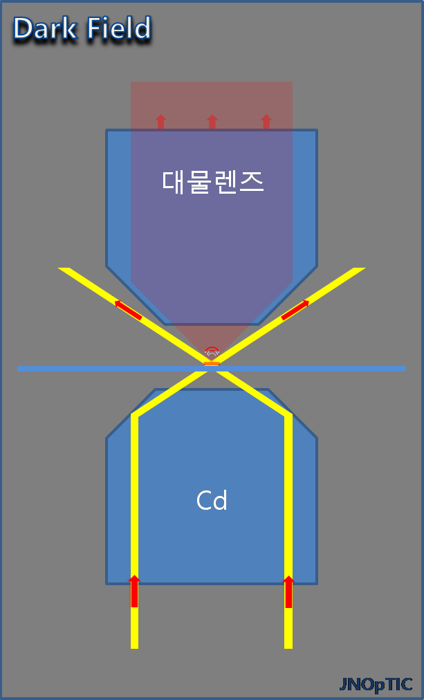
Dark Field

암시야 현미경 관찰법

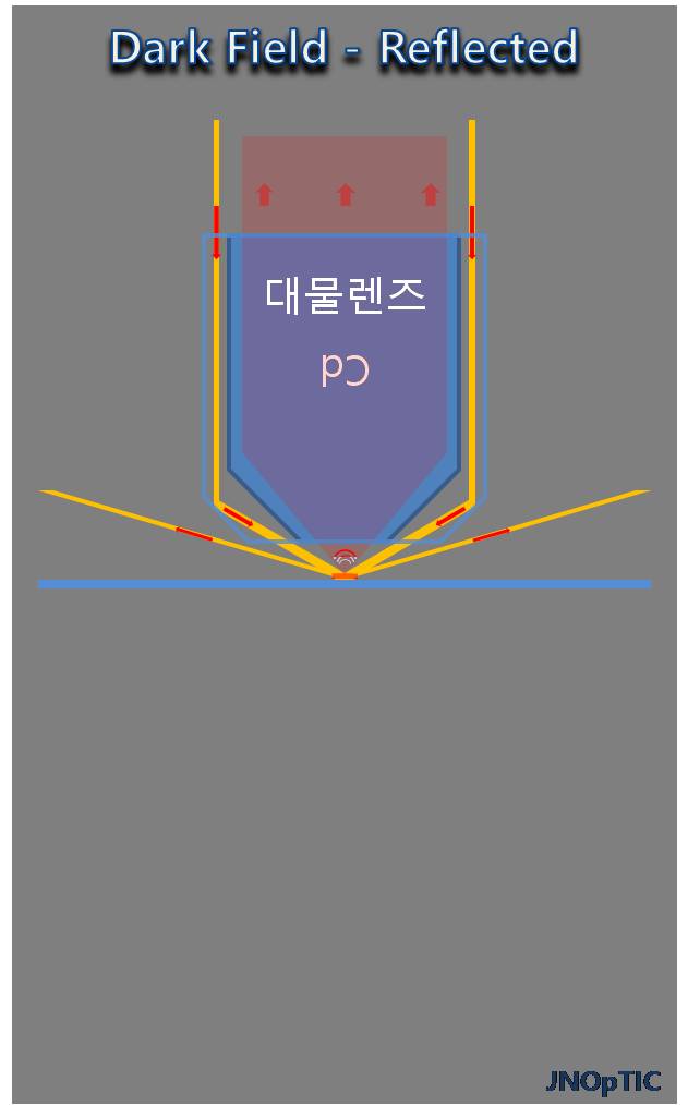
Dark Field Microscopy (Transmitted & Reflected)
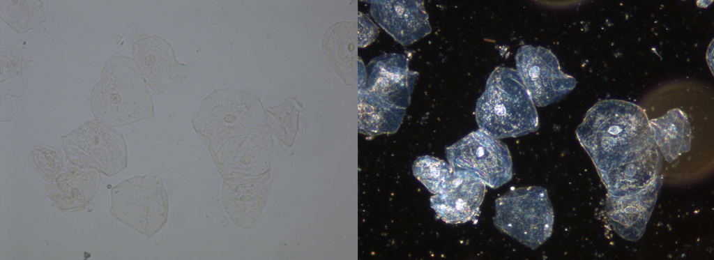
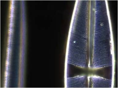
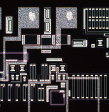
명시야(BF) 이미지와 암시야(DF) 이미지의 비교
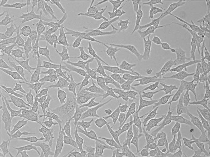
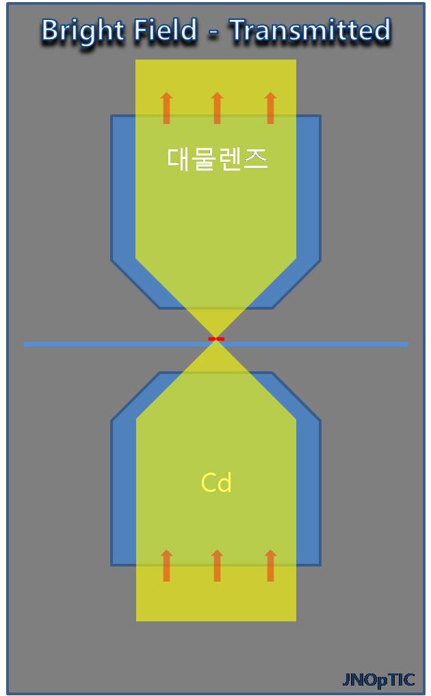
투과 현미경에서 보는 Bright filed Image & Dark Field Image

암시야 (Dark field ) 검경법 의 특징

  • 스크래치나 단차, 파티클을 강조하는 관찰법
  • 샘플에 닿는 직접 조명광은 밖으로 유도하고, 오로지 샘플에서 나오는 산란광 만을 대물렌즈로 취하는 관찰법
  • 배경이 어두운 상태에서 샘플이 반짝이기 때문에 현미경의 분해능을 넘어서는 관찰이 가능하다. 

생물 (Bio) 현미경

투과 조명 _ Bright Field 와 Dark Field 

산업(금속) 현미경

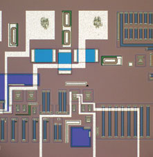
반사조명 _ Bright Field 와 Dark Field 

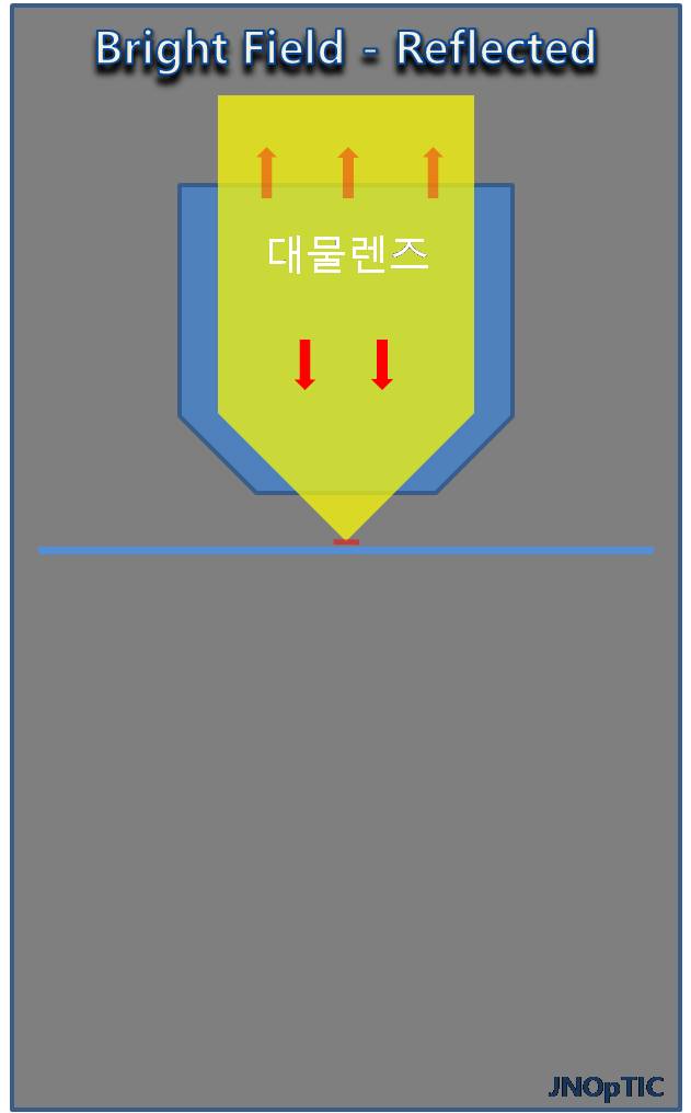
Bright Field

명시야 현미경 관찰법 설명

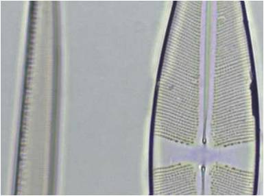
Bright Field Microscopy

반사조명이미지
반사조명 이미지

Bright Field 관찰법 발명자 정보

  Bright Field 관찰법은 현미경 관찰의 시작이라고 할 수 있습니다. 샘플을 확대하는 등의 여러 발명은 아주 오랜 전 부터 기록을 볼 수 있었으며,  1590년대의 네덜란드인 얀센 (Zacharias Jansen)이 현재의 현미경과 같은 구조의 현미경을 제작하였고, 그 이후로 현대에 이르기까지 다양한 관찰법의 시작점으로 사용되어 지고 있습니다.  

Bright Field 현미경의 개요

가장 기본이 되는 관찰법. 
금속 또는 산업현미경
  • 대부분의 샘플이 불투명하여 반사조명을 주로 사용
생물 현미경
  • 대부분의 샘플이 투명하여 투과조명을 주로 사용 

필요에 따라서 반사와 투과 조명을 동시에 사용하는 경우도 있음.

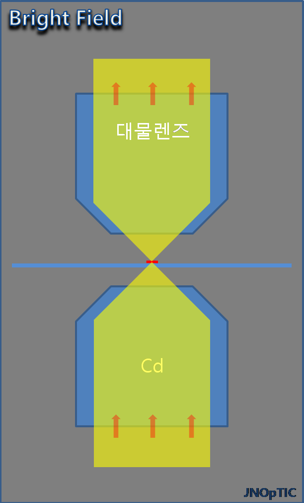
반사 현미경과 투과현미경에서 조명 차이

  • Bright Field 현미경의 설명

샘플을 단순 확대 관찰 용도이며,  특수한 광학유닛을 필요로 하지 않는 기본 관찰 방법입니다. 현미경에서 사용되는 조명계는 과거에는 Critical illumination을 사용하였으나, 현재에 판매되는 대부분의 현미경의 조명계는 쾰러 일루미네이션(Köhler illumination )을 사용하고 있습니다.

 조명에 의해 만들어 지는 결상계와 샘플에 의해 만들어지는 결상계가 서로 영향을 미치지 않도록 설계되어 있으며, 조명의 결상에 의해 발생하는 샘플의 이미지 저하를 최소화 한 조명계입니다.

critical illumination : 예전에 사용하였던 조명 방법으로 조명의 결상계과 샘플의 결상계가 서로 중복 결상되도록 되어 있어서, 샘플의 관찰을 할 때 조명의 상과 샘플의 상이 중복되어 관찰되어 이미지의 저하가 발생합니다.  

수차(aberration)의 종류

자이델의 5수차 (The Five Seidel Aberrations )
  • Spherical Aberration ( 구면수차 )
  • Coma ( 혜성형 수차 )
  • Astigmatism ( 비점수차 )
  • Curvature of field ( 상면만곡 ) _ 대물렌즈의 보정 표시 ( PLAN )
  • Distortion ( 왜곡 )
색수차 ( Chromatic aberration )

가시광선 대역에 있어서 파장이 짧은 보라색의 빛과 파장이 긴 붉은색의 빛의 굴절율을 보면 파장이 짧을 수록 렌즈를 통과한 후의 굴절율이 크고, 파장이 길수록 굴절율이 작아지기 때문에 하나의 렌즈로 빛을 결상하고자 할때 파장에 따라 결상 위치에 차이를 발생하게 됩니다. 일반적으로 현미경에서 사용되는 대물렌즈는 이러한 색수차에 의한 문제점을 보정 하는 정도로 다음과 같이 대물렌즈의 등급을 분류하고 있습니다.

  • Achromat – 저가형 현미경에 주로 사용되어지는 렌즈
  • Flourite ( Semi-Apo ) – 현업에서 많이 사용되는 범용 렌즈
  • Apochromat – 고급 렌즈로 가격이 가장 비싼 렌즈

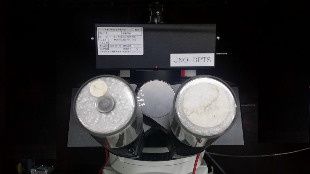
JNO-DPTS

Multi-Dual Port ( Simultaneously Observation )

Download  Catalog  JNO-DPTS

To observe Fluorescence image  and IR DIC image  at the same time
with Multi-Dual Port tube for BX51-WI
(Made by Jae Hwan JIN )

JNOPTIC Multi-Dual Port System
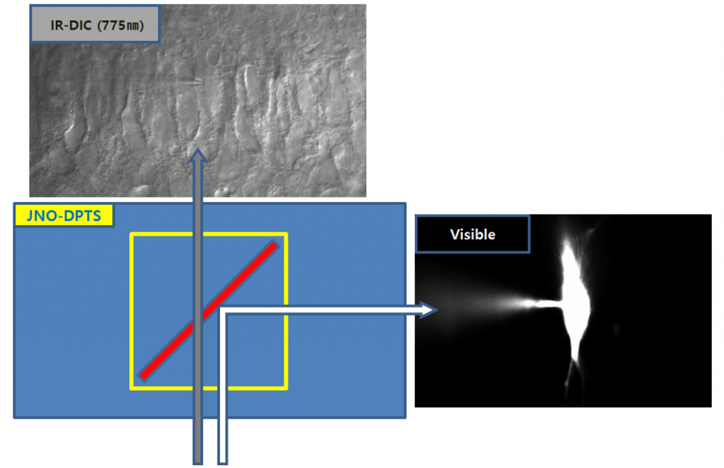
  1. Simultaneously Observation
Simultaneously Observation
IR-DIC 이미지는 투과 되고, Visivle 이미지 반사되어 각각의 카메라로 전달 된다.

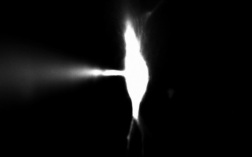
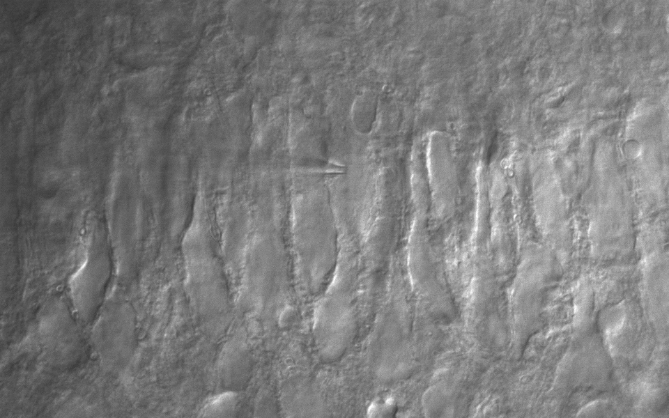
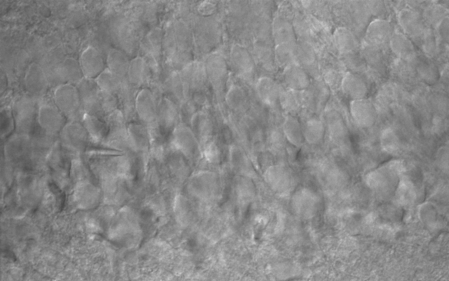
CA3 pyramidal neuron, IR-DIC & Alexa Fluor 488 (50 uM)

20160323_1(FullF6)_2 for wp
20160323_1(1) for wp
CA3 pyramidal neuron Alexa Fluor 488, IR-DIC (Simultaneous observations)
Figure courtesy by Dr. Sooyun Kim, Seoul National University
20151209_2_stim1(CA3 pyramidal neuron) for wp
CA3 pyramidal neuron
AcquCAM 23S2 & JNO-RNB filter with BX51WI
20151209_2(CA3 pyramidal neuron) for wp
CA3 pyramidal neuron
AcquCAM 23S2 & JNO-RNB filter with BX51WI
Frame Rate162 fps, increased through ROI
Sensor1 / 1.2 inch ( 1920×1200, 5.86㎛ x 5.86㎛ )
FILTERJNO-RemN/B (Noisy cut filter)
Manification1x, 1.25x, 1.6x, 2x

JNO-ARM

Advance Realtime Monitor. Easy-to-Use, Image Enhancement, etc

1. Information

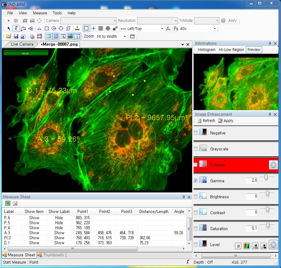
ARM is image analysis software for JNOPTIC AcquCAM cameras. This S/W is interchangeable with all WDM cameras regardless of camera brand and model and even more the most strength point is simple and easy use of length, area, angle, etc.ARM01
2. Simple measurement tools

toolbar

  • Measurement Tools: Count, Distance, Angle, Area, etc.
  • Guide Lines: Cross Line, Rectangular Lattice, Circular Grid
  • Scale Panel on Live/Image Screen
  • Available measurement on LIVE image and saved image mode

3-1. Specialization for observation of fluorescence images

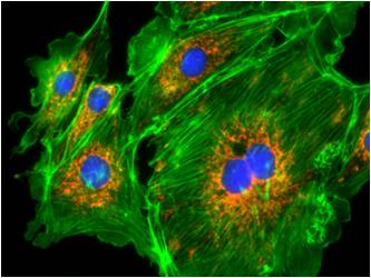
Improvement Function about Fluorescence Microscopy Image. (Used function: Auto Level, Low level, Pseudo Color, Merge Image)
ARM02
 Bottom on the left is merged image without improvement of image. And the bottom on the right is merged image after improvement of image. These images are shot by JNOPTIC AcquCAM Pro/G3 camera

3-2-1 Effect of high-contrast image with simple operation (Monochrome)

Improvement Function about Microscopy Image Monochrome. (Used function: Auto Level)arm04Effective improvement of image with simple operation. This image is shot by JNOPTIC Pro/S3 camera

3-2-2 Effect of high-contrast image with simple operation (Color)

Improvement Function about Microscopy Image. (Used function: Auto Level)arm05Effective improvement of image with simple operation. These images are shot by JNOPTIC AcquCAM Pro/G3 camera

3-3-1 Live Pseudo Color (for color camera)

When you get fluorescence images using color camera, if the unwanted channel was shown because of cross talk, you could remove this channel to take advantage of function of Live Pseudo.
Improvement Function about Fluorescence Microscopy Live Camera Image. (Used function: Live Pseudo Color)
arm06
 The extraction of wanted color information from color image (Remove BR on RGB source) These images are shot by JNOPTIC AcquCAM Pro/G3 camera

3-3-2 Live Pseudo Color (for Monochrome camera)

When you get fluorescence images using monochrome camera, you can raise efficiency of acquisition for fluorescence images to add similar false color to the color to be shown on the eyepiece in real time.
Improvement Function about Fluorescence Microscopy Live Monochrome Camera Image. (Used function: Live Pseudo Color)
arm08
 Improvement of usage environment for monochrome camera to add false color on the image. These images are shot by JNOPTIC AcquCAM Pro/S3 camera

4. JNO-MHU(Option Unit)

Available measuring height with additional unit, JNO-MHU
※ JNO-MHU is sold separately as optional unit.
jno-mhu1
  jno-mhu2
 Left: Focus on top of sample (Z-axis reset), Right: Focus on bottom of sample (Z-axis height measurement)
jno-mhu3
 < Measuring condition : over 20x objective, temp 20°C >
Responding Model Measurement value unit Measurable height Error of measuring 1000㎛ Recommended measuring height
CX 41 0.2 ㎛ -9.9 ~ 29.9㎜ Below ±20㎛ Below ± 2000㎛
CKX 41 0.2 ㎛ -9.9 ~ 29.9㎜ Below ±20㎛ Below ± 2000㎛
BX – FM 0.2 ㎛ -9.9 ~ 29.9㎜ Below ±20㎛ Below ± 2000㎛
BX 51/53 0.1 ㎛ -9.9 ~ 29.9㎜ Below ±10㎛ Below ± 1000㎛
MX 51 0.1 ㎛ -9.9 ~ 29.9㎜ Below ±10㎛ Below ± 1000㎛
MX 61L/61 0.1 ㎛ -9.9 ~ 29.9㎜ Below ±10㎛ Below ± 1000㎛

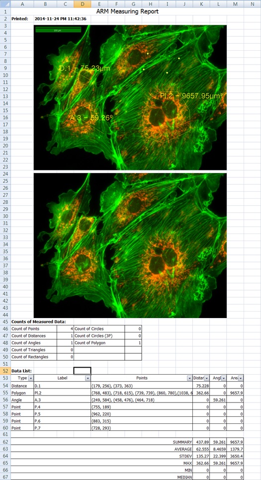
5. Reporting to Excel
excel


OpTIC Eye (Material)

JNOPTIC Image Analysis Software OpTIC Eye Series

Catalog_OpTIC Eye

  1. OpTIC Eye (Material)

기본측정+오토카운팅(입도분석용)+금속조직분석용
– 형상측정(상분율측정)
: 두가지 이상의 색상을 추출하여 면적율 계산
– 그레인사이즈(Grain Size Analysis)
: ASTM E 1382에 의한 금속표면의 결정립도 산출
: 교차점 / 면적 / 수동면적 추출방식
– 구상화율 분석(Nodular-graphite analysis)
: ASTM, ISO, JIS 규격에 따라 분석이 가능
– 비금속 개재물(Non-Metallic Inclusion)
: 산화물, 규산물, 황화물, 내화물, 광재등 추출


영상분석에 의한 구상화율 측정 (nodular-graphite analysis)

주철분석 소프트웨어 모듈로 ASTM, JIS, KS 규격에 따라 구상흑연주철의 구상흑연에 대하여 현미경영상분석을 통해 구상화율을 측정하고 결과를 표시합니다.

20
구상화율 측정 모듈의 사용자 인터페이스


영상분석에 의한 결정도립 측정 (rain Size Analysis)


  • ASTM에 의거, 금속표면의 Grainsize(결정립크기) 및 ASTM Grain Size Number(결정립도)를 산출합니다.
  • ASTM 결정립도를산출하기 위하여 교차추출 및 면적추출방식중 택일하여 간단히 측정할 수 있습니다.

22
교차추출(Intercept)방식의 측정

23
면적추출(Planimetric)방식의 측정


OpTIC Eye (Auto)

JNOPTIC Image Analysis Software OpTIC Eye Series

Catalog_OpTIC Eye

  1. OpTIC Eye (Auto)

기본측정+보고서+오토카운팅(입도분석용)
– 셀이미지 자동측정(5단계 easy측정)
: 영상분할, 측정항목설정, 측정, 분류, 성적서기능
– 면적, 면적비, 반경, 직경등 15개 항목 자동측정

Auto Counting

영상을 자동 분석하는 기능입니다. 영상을 개체들(Objects)과  배경(Background)으로 분할(Segmentation)함 으로써 측정할 영역을 한번에 검출해내고, 면적 등을 자동측정 합니다. Auto Count는 위의 차례대로 측정이 이루어 집니다.

  • 영상분할(Segmentation)
  • 측정항목설정(Define Measure)
  • 측정(Measure)
  • 분류(Classification)
  • 성적서(Report)

측정 항목 설정(Define Measure)

1

  • 원하는 측정 항목 설정
  • 측정항목별 그림에 의한 상세설명
  • 구간 설정: 상한, 하한 적용가능

측정결과를 분석/분류 (Classification)

23

View Measurement를 통한 실시간 측정 항목 display 측정

크기별로 그룹화(Classification), 그룹별 색상화 가능

Report

4

측정결과를 4가지 형태의 Chart로 표시: Histogram, Line graph, Pie, Scatter-gram Excel 전송 가능

JNO-WI-3

X Y Mover & Bridge Stage

Description
Material   Aluminum
표면 처리   Black –Anodizing
Travel Range (mm)   X    ±12.5     Y    ±12.5
높이 (mm)   170  ~ 220
Tap Hole 규격   M6
Tap Hole 간격 (mm)   X    25    Y    25


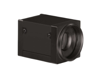
AcquCAM 23S

AcquCAM 23S

(Down load Catalog ACQUCAM 23S)

  • USB 3.0 Monochrome Camera
  • 1/1.2 ” Exmor Sensor
  • 1920×1200 pixel
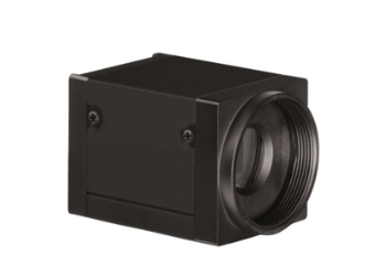
  • Trigger input and I/O

AcquCAM 23S
EXMOR Sensors: Next-generation imaging sensors based on CMOS, increase the strength and reduce noise.


High sensitivity camera available from visible to IR range

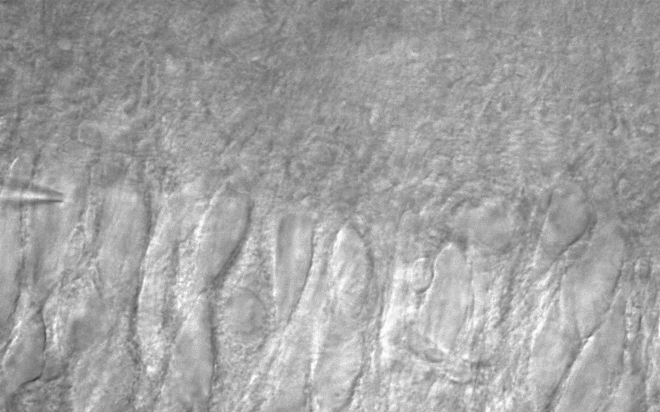
20151120_1(CA1 pyramidal neuron)
Apply high sensitivity EXMOR sensor with additional filter block visible range, IR Cut filter image could be observed.


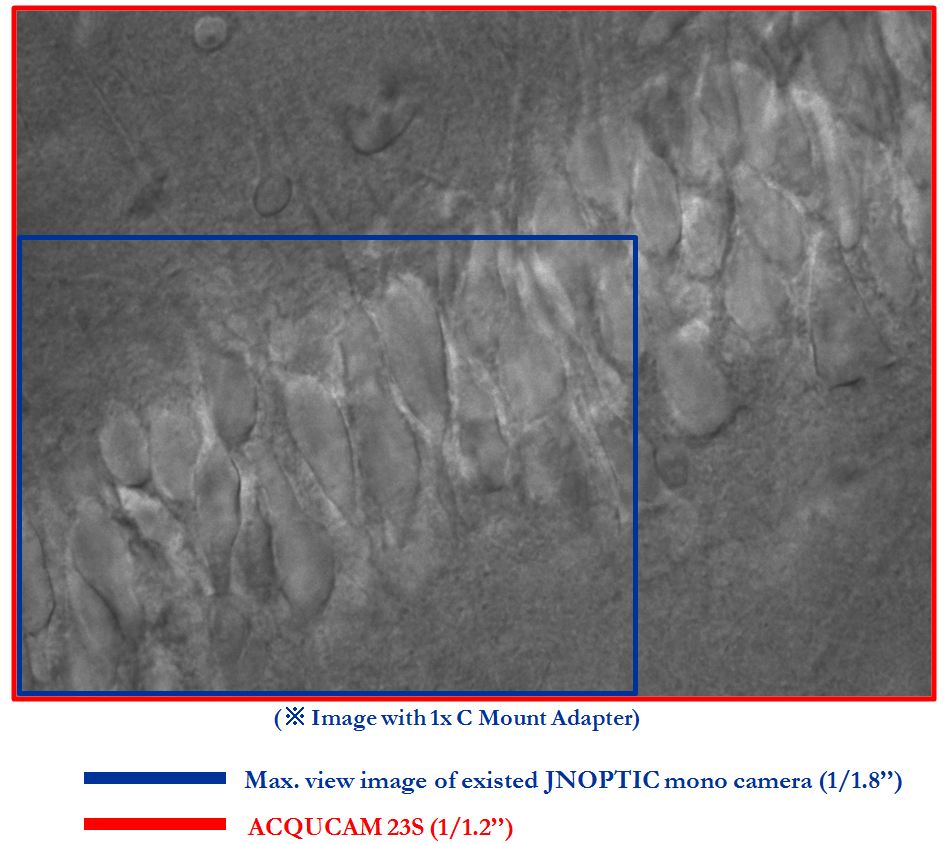
Available wider range of observation

AcquCAM 23S_6
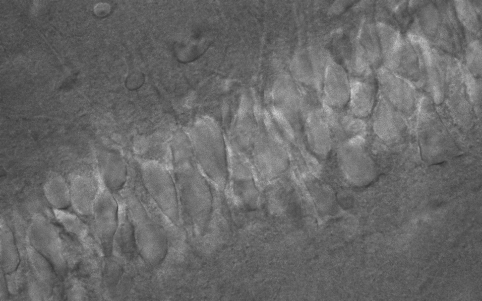
By applying a 1 / 1.2 ” EXMOR sensor, you can observe a wider range of superior sensitivity image.
These images were shot by AcquCAM 23S Package.

High sensitivity and fast frame image

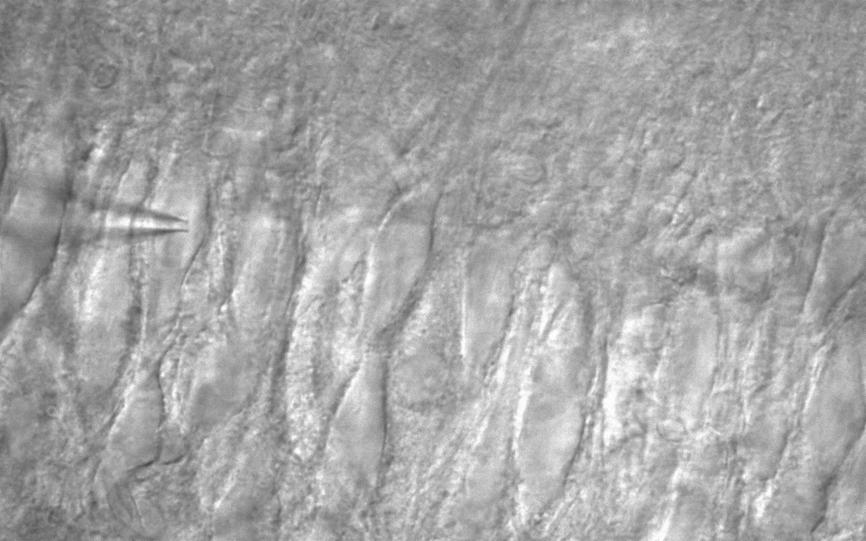
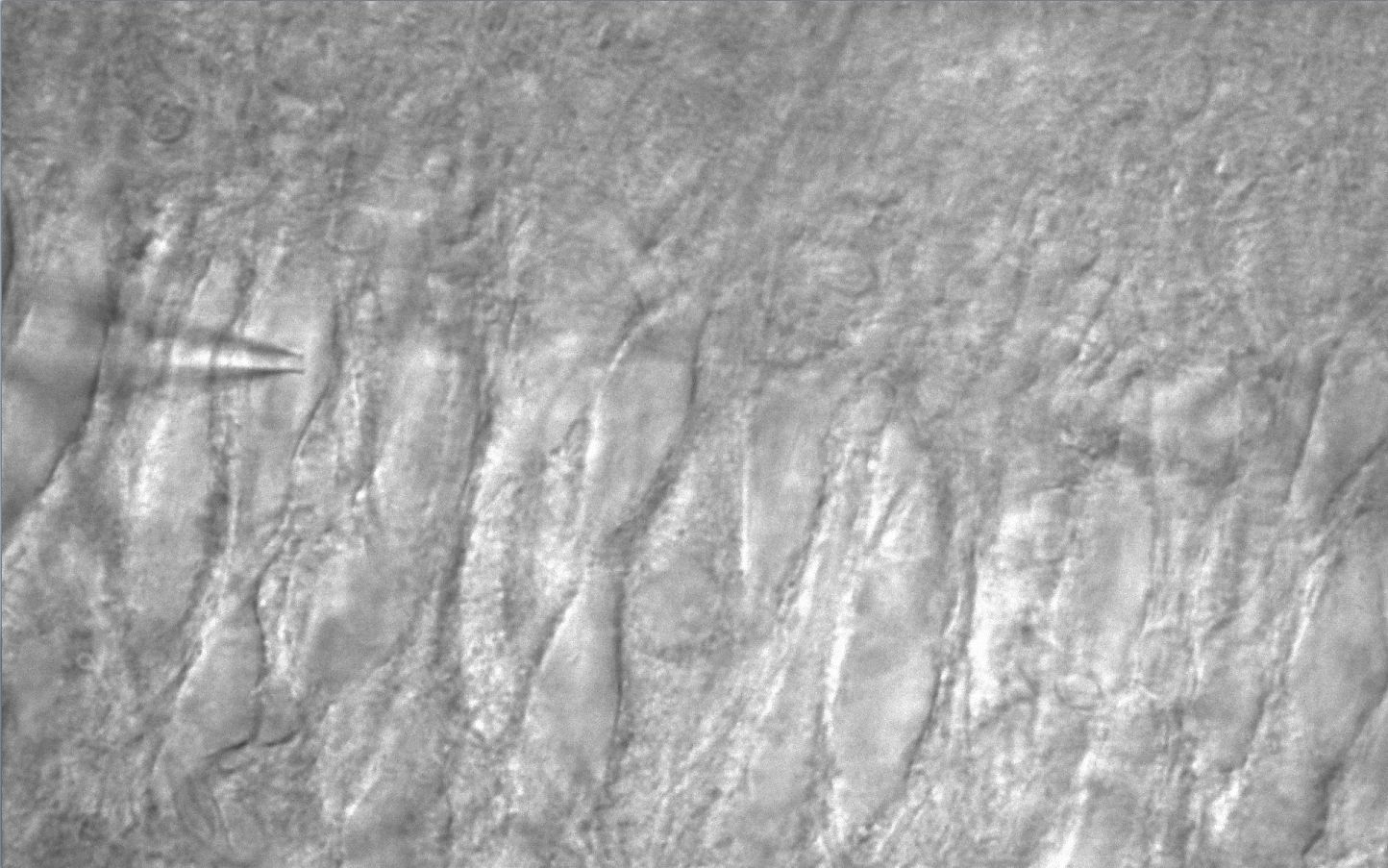
AcquCAM 23S_(CA3 pyramidal neuron)

AcquCAM 23S_(Dentate gyrus Granule cell)
To maximize the range of imaging possible observation with fast frame 54Fps and high sensitivity 0.015 lx .

[Specification]

Spec Description
Size 1/1.2 inch
Resolution H: 1920, V: 1200
Pixel size H: 5.86 µm, V: 5.86 µm
Video formats

& Frame rate

1920×1200 Y16 @ 25 fps

1920×1200 Y800 @ 54 fps

Sensitivity 0.015 lx
Exposure time 1/100000 to 30 s
Lens mount C/Cs mount
Interface USB 3.0
Power supply 4.5 to 5.5 VDC
Dimensions H: 29 mm, W: 29 mm, L: 43 mm
Mass 65 g


AcquCAM Pro/S3

AcquCAM Pro/S3

(Down load Catalog_ACQUCAM PRO/S3)

AcquCAM Pro/S3

AcquCAM Pro/S3 USB 3.0 camera is the perfect solution for many bio, industrial, medical applications. Utilizing the very sensitive mono CCD sensors, these cameras feature a specialization of fluorescence observations. With up to 30 fps and a trigger delay of less than 5 micro seconds.

  • 1.2 Mega pixel CCDcamera
  • Monochrome, Progressive scan
  • Preview software provided (JNOPTIC Capture)
  • Software compatible with Windows XP/ Vista/ 7/ 8 operating systems

Apply Pseudo color for live fluorescence

Apply Pseudo color for black live fluorescence

AcquCAM Pro S3_6

Emphasis function for variety images

Getting clear image with general light source

[Specification]

SpecDescription
Size1/3 inch
ResolutionH: 1280, V: 960
Pixel sizeH: 3.75 µm, V: 3.75 µm
Video formats & Frame rate1280×960 Y800 @ 30, 20, 15, 7.5, 3.75 fps 1280×960 Y16 @ 30, 20, 15, 7.5, 3.75 fps
Sensitivity0.015 lx
Exposure time1/10000 to 30 s
Lens mountC/Cs mount
InterfaceUSB 3.0
Power supply4.5 to 5.5 VDC
DimensionsH: 29 mm, W: 29 mm, L: 43 mm
Mass65 g