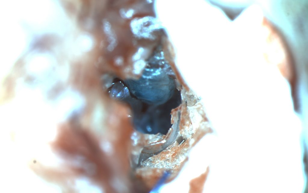
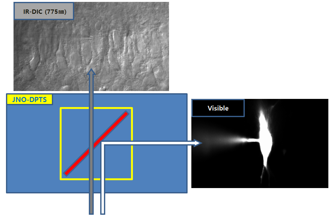
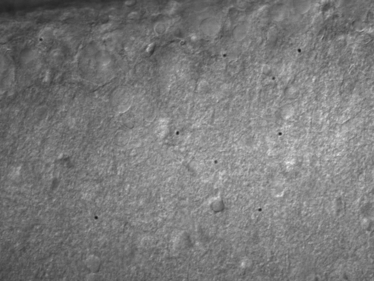
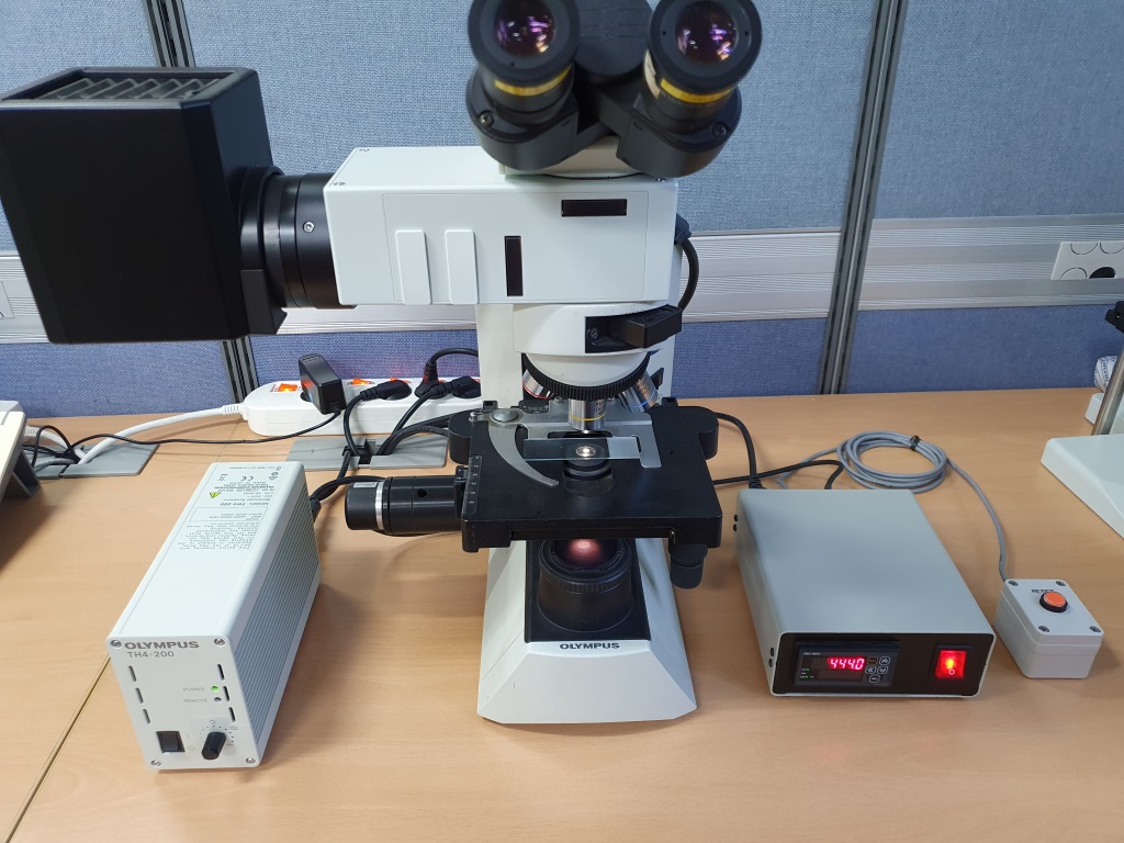
CA3 pyramidal neuron IR-DIC
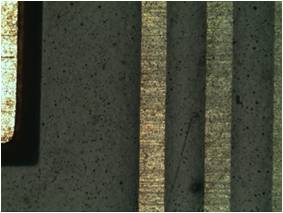
Figure courtesy by Dr. Sooyun Kim, Seoul National UniversityFilter Set : JNO-B(B)
Objective Lens : PlanApo60xWLSMJNO – DPTS Made by JinOpticDIC Upgrade Image by J.H.JIN
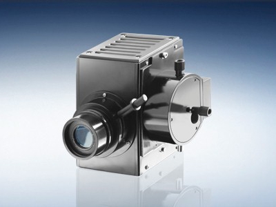
Microscope : NIKON FN1 & JNO-GGE
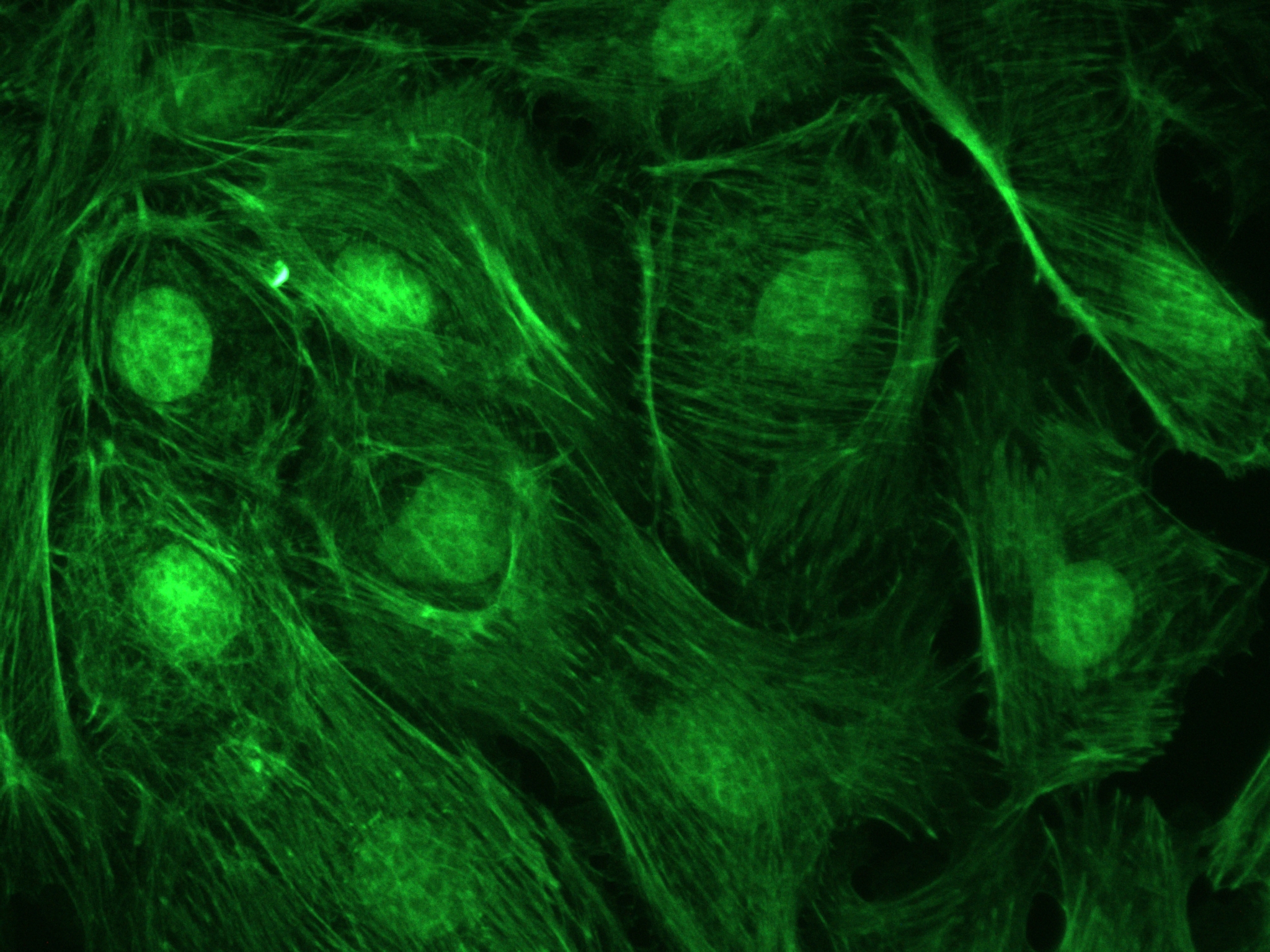
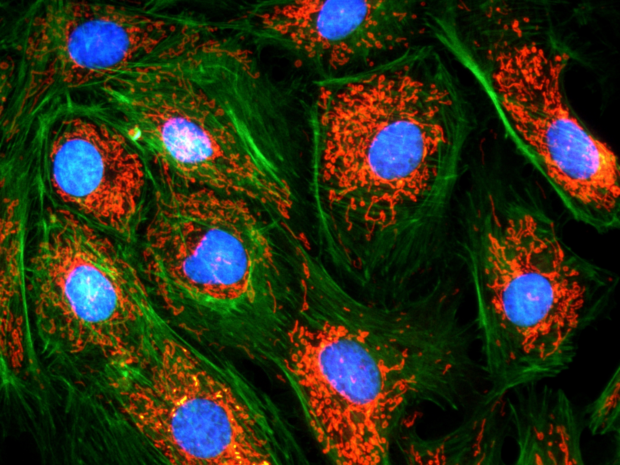
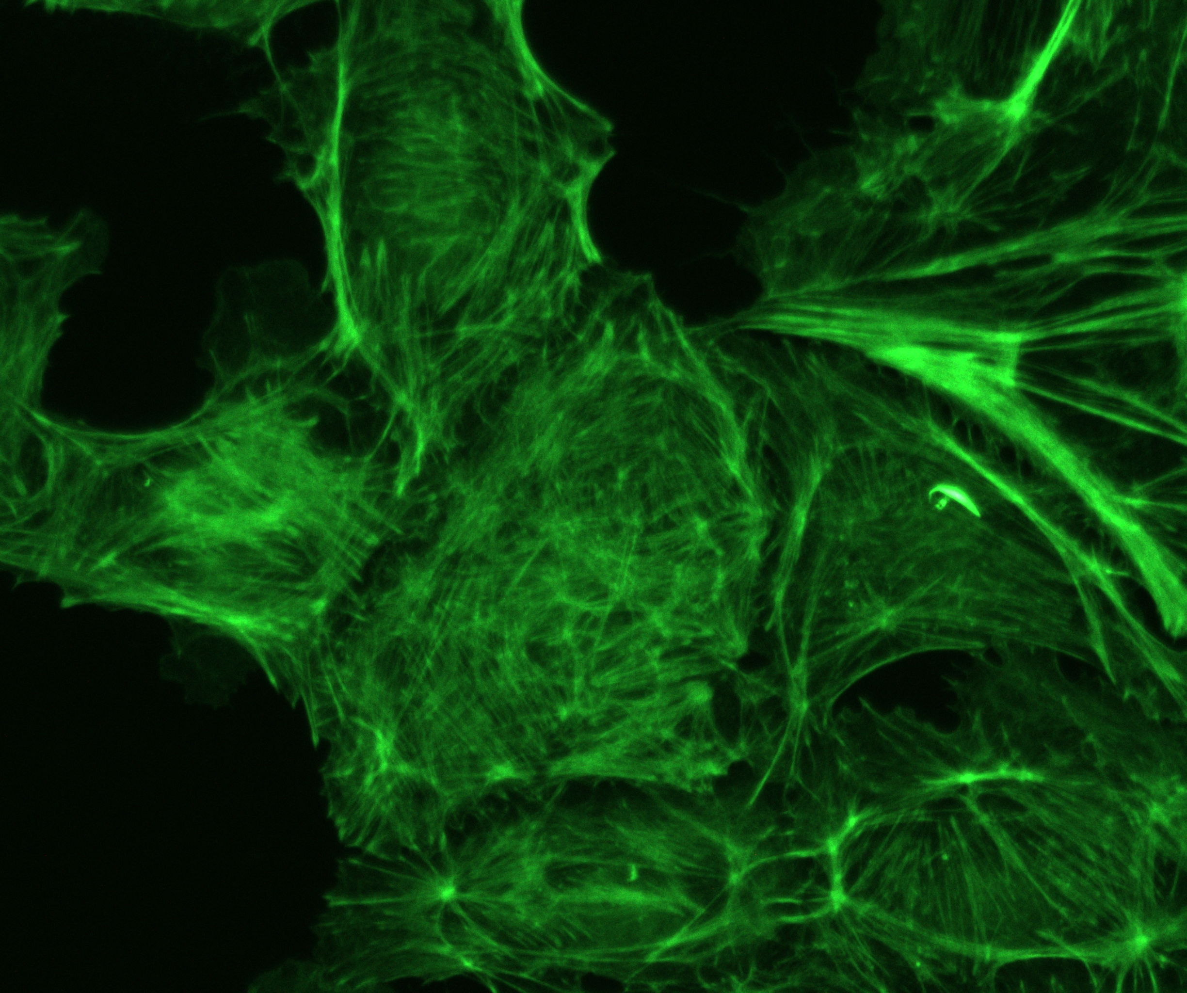
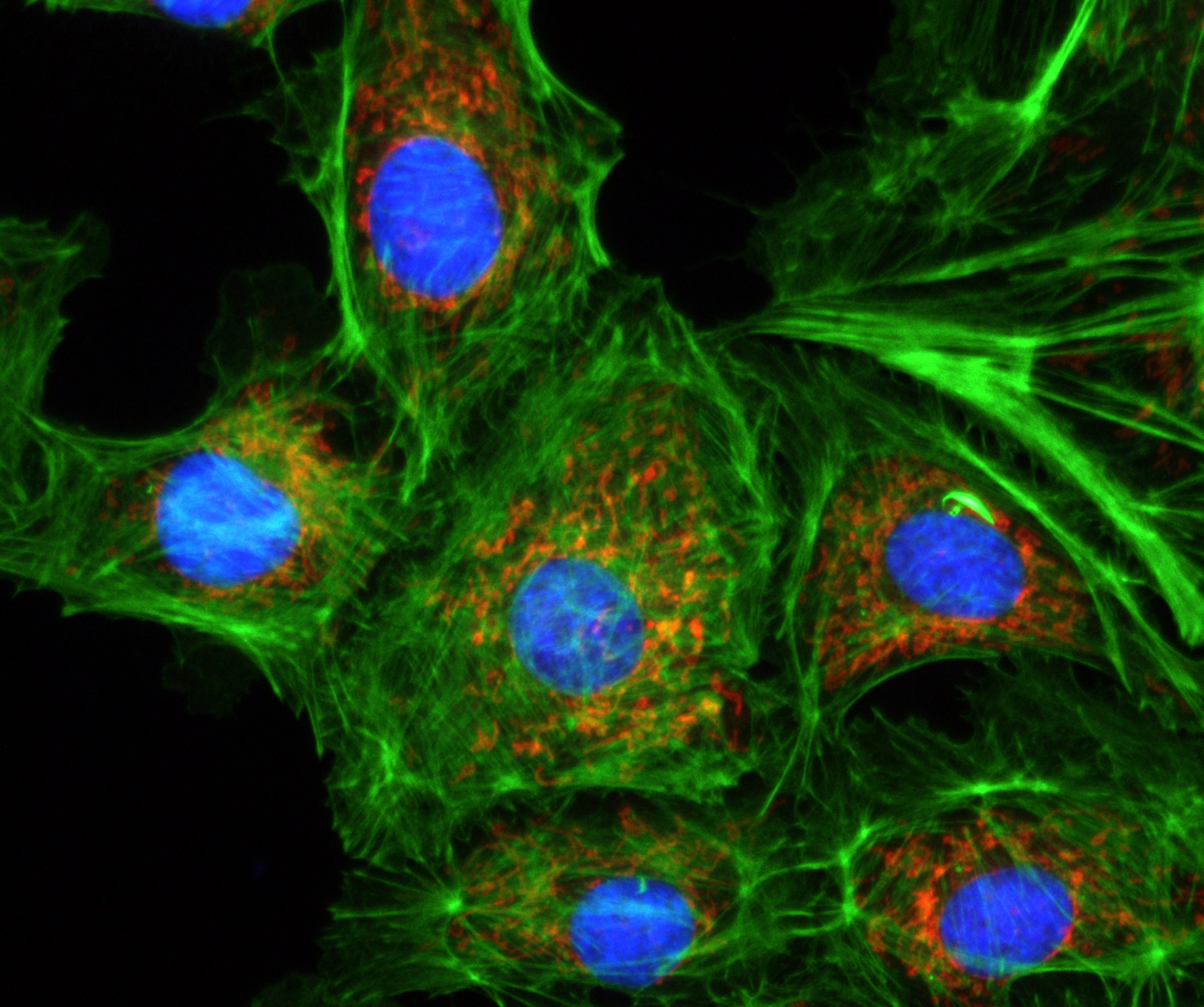
Cyanines were and are still used in industry, and more recently in biotechnology (labeling, analysis). Cyanines have many uses as fluorescent dyes, particularly in biomedical imaging. Depending on the structure, they cover the spectrum from IR to UV.
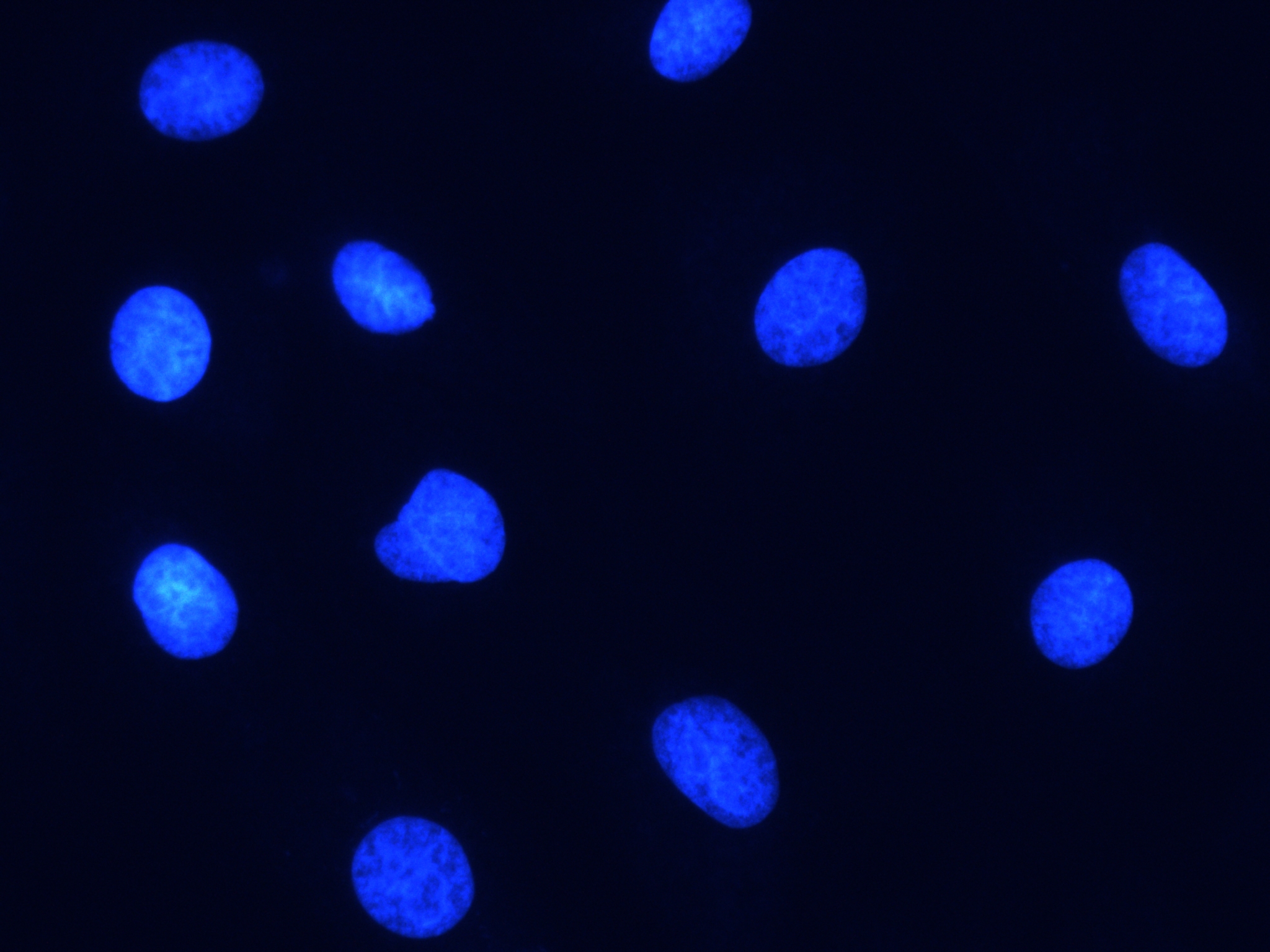
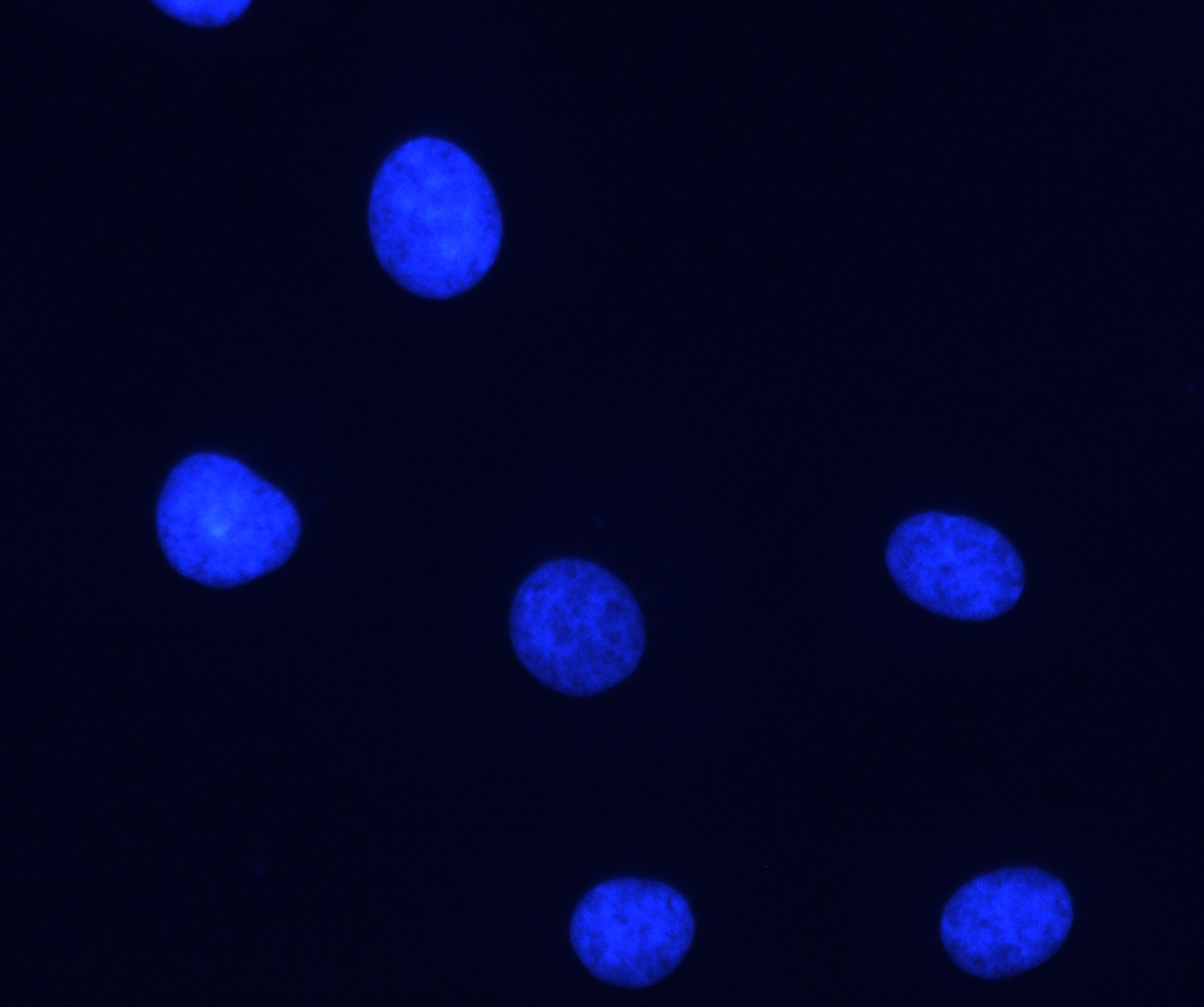
Cyanine is the non-systematic name of a synthetic dye family belonging to polymethine group. The word cyanin is from the English word “cyan”, which conventionally means a shade of blue-green (close to “aqua”) and is derived from the Greek κυάνεος/κυανοῦς kyaneos/kyanous which means a somewhat different color: “dark blue”.
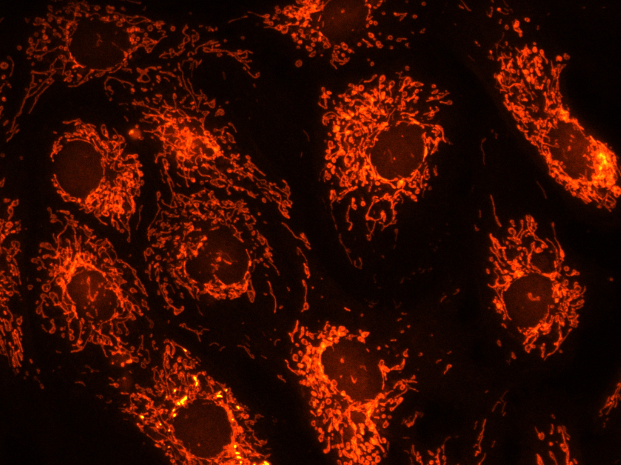
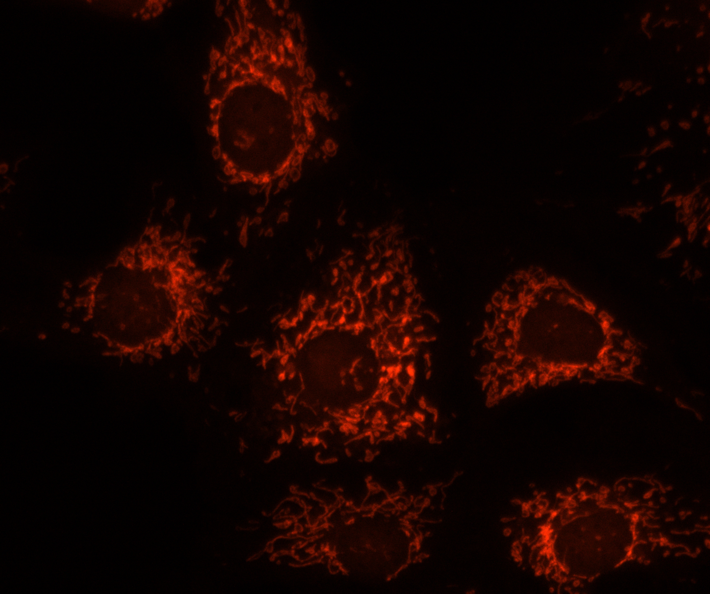
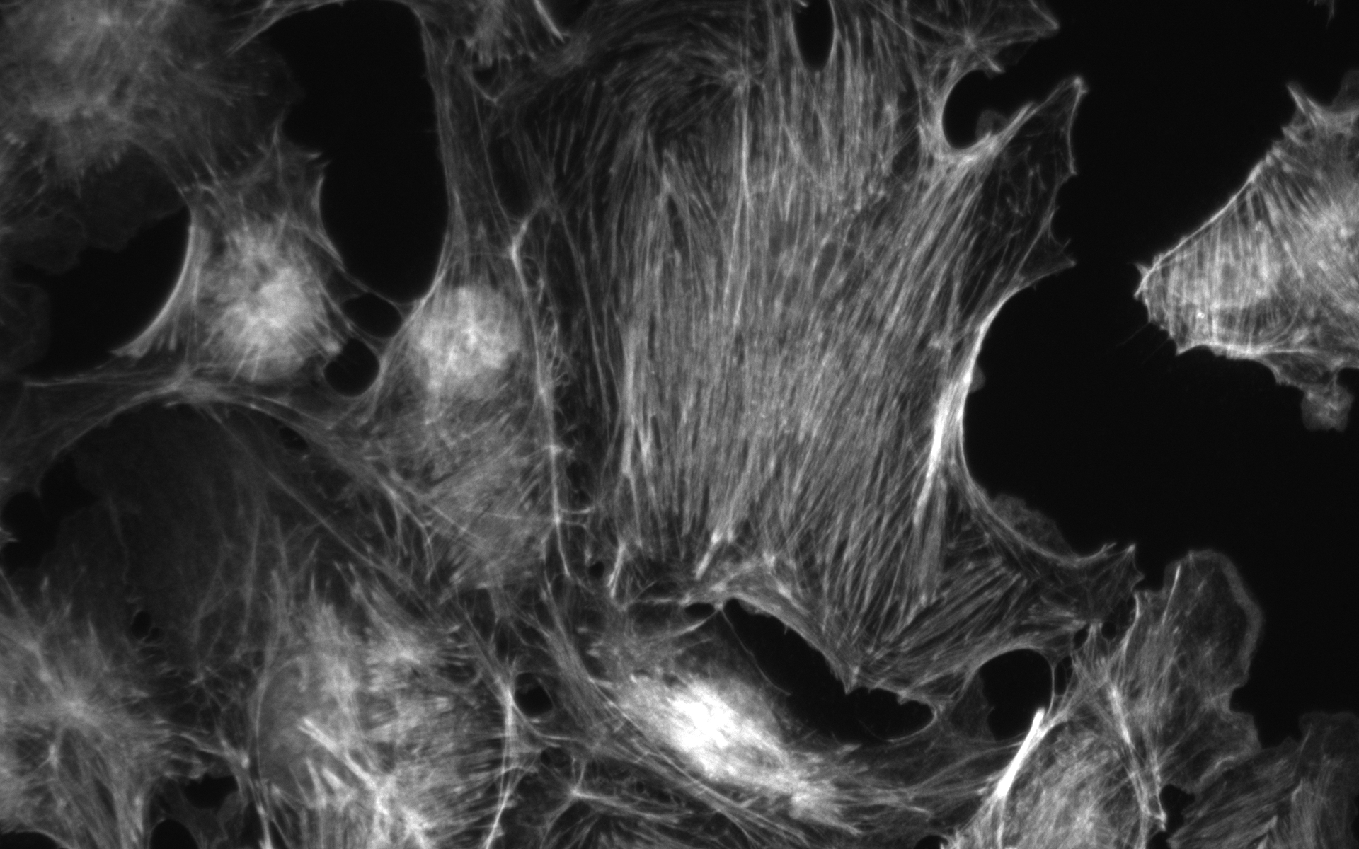
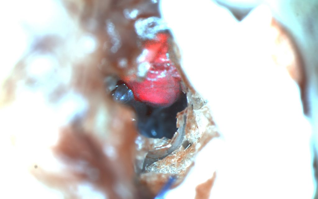
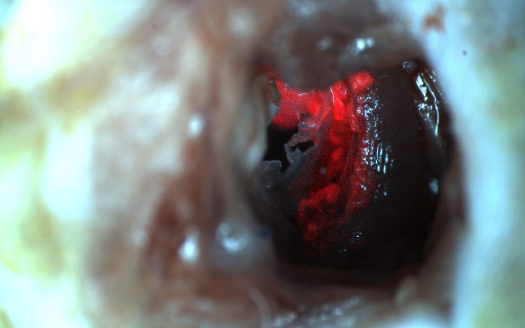
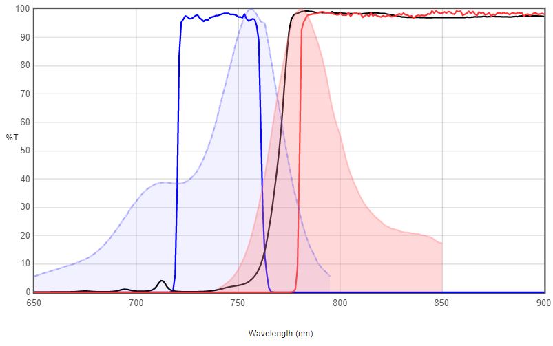
Cy7 is a near-IR fluor that is invisible to the naked eye (Excitation/emission maximum 750/776 ㎚). It is used in in vivo imaging applications, as well as the Cy7.5 dye.
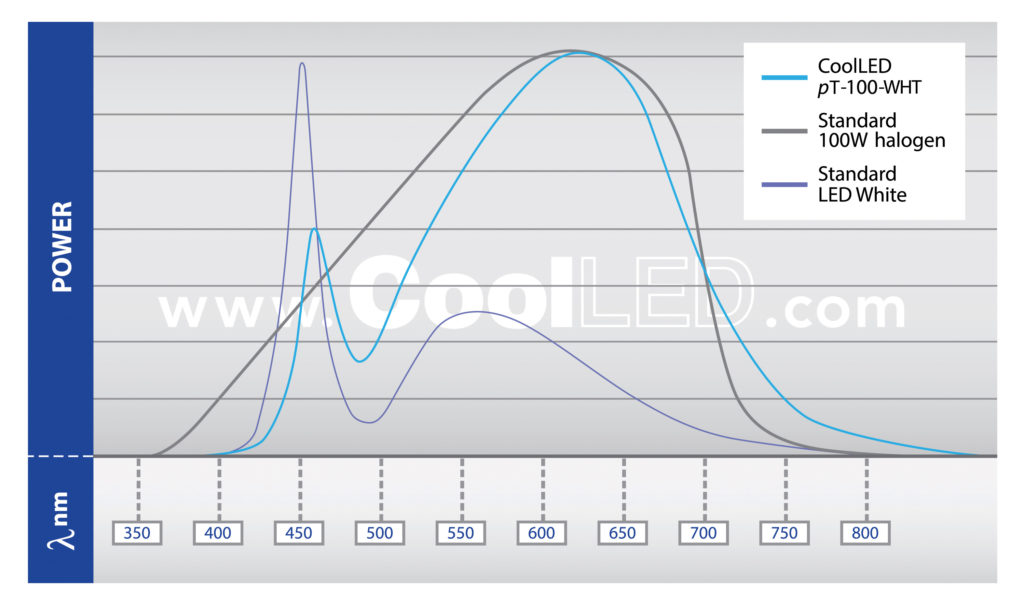
Cyanines(Cy) replace advantageously conventional dyes such as Fluorescein(FITC) and rhodamines (TRITC, RRX), yielding brighter and more stable fluorescence.
시아닌은 여전히 업계에서, 그리고 최근에는 생명 공학 (라벨링, 분석)에서 사용되고 있습니다. 시아닌은 형광 염료로서, 특히 생체 의학 이미징에서 많은 용도로 사용되며 구조에 따라 IR에서 UV까지 스펙트럼을 커버합니다.
Cyanine은 polymethine 그룹에 속하는 합성 염료 계열의 비 계통적 이름입니다. cyanin이라는 단어는 일반적으로 청록색 ( “아쿠아”에 가까운)의 그늘을 의미하는 영어 단어 “cyan”에서 유래되었으며 다소 다른 색상 “진한 파란색 “을 의미하는 그리스어 κυάνεος / κυανοῦς kyaneos / kyanous에서 파생되었습니다.
Cy7은 육안으로는 보이지 않는 근적외선 형광체이며, Cy7.5 염료와 함께 생체 내 이미징 어플리케이션에 사용됩니다.
Cyanines (Cy)는 Fluorescein (FITC) 및 rhodamines (TRITC, RRX)와 같은 기존의 염료를 대체하여 보다 밝고 안정한 형광을 생성합니다.