jhjin의 모든 글

AcquCAM Pro/G3

AcquCAM Pro/G3

Down load Catalog _ AcquCAM PRO/G3

AcquCAM Pro/G3

 AcquCAM Pro/G3 USB 3.0 camera is the perfect solution for many bio, industrial, medical applications. Utilizing the very sensitive color CCD sensors, these cameras feature a specialization of fluorescence observations. With up to 30 fps and a trigger delay of less than 5 micro seconds.

  • 1.2 Mega pixel CCD camera
  • Color, Progressive scan
  • Preview software provided (JNOPTIC Capture)
  • Software compatible with Windows XP/ Vista/ 7/ 8 operating systems

LIVE PSEUDO COLOR

Ex. of applied PSEUDO COLOR for black live fluorescence image

Emphasis function on variety of images

AcquCAM Pro G3_15
With emphasis function, BLACK BALANCE / AUTO LEVEL of ARM on variety images, you could acquire cleaner and sharper images.

Acquiring high sensitive images

These images were shot by AcquCAM Silver Package.

Acquiring high sensitive images from general lighting source

[Specification]

SpecDescription
Size1/3 inch
ResolutionH: 1280, V: 960
Pixel sizeH: 3.75 µm, V: 3.75 µm
Video formats   & Frame rate1280×960 RGB32 @ 30, 20, 15, 7.5, 3.75 fps   1280×960 Y800 @ 30, 20, 15, 7.5, 3.75 fps 1280×960 Y16 @ 30, 20, 15, 7.5, 3.75 fps
Sensitivity0.05 lx
Exposure time1/10000 to 30 s
Lens mountC/Cs mount
InterfaceUSB 3.0
Power supply4.5 to 5.5 VDC
DimensionsH: 29 mm, W: 29 mm, L: 43 mm
Mass65 g

AcquCAM 23S2

AcquCAM 23S2

(Down load Catalog – AcquCAM 23S2)

CA3 pyramidal neuron, IR-DIC & Alexa Fluo 488 at the same time
Figure courtesy by Dr. Sooyun Kim, Seoul National University
with Bx51WI, JNO-DPTS, AcquCAM 23S
JNO-DPTS
CA3 pyramidal neuron, IR-DIC & Alexa Fluo 488 at the same time
Figure courtesy by Dr. Sooyun Kim, Seoul National University
with Bx51WI, JNO-DPTS, AcquCAM 23S
  • USB 3.0 Monochrome Camera
  • 1/1.2 ” Pregius Sensor
  • 1920×1200 pixel
  • Trigger input and I/O
  • Cost-effective mono camera for fluorescence microscope (Best)
  • Cost-effective camera for Department of Physiology (Best)
  • 1/1.2 inch optimized sensor size for microscope field of view
  • Acquire images of optimum width combined with 0.63x adapter

AcquCAM 23S2 mono camera has a USB 3.0 interface and is the perfect solution for many industrial automation, quality assurance, security, surveillance and medical applications. The mono camera ships with the very sensitive 1/1.2 inch CMOS Pregius  sensor. With up to 162 images per second, the AcuqCAM 23S2 is a low cost, yet highly versatile imaging solution. The camera includes a C to CS mount adapter, making it compatible to C and CS mount lenses.

Images with AcquCAM 23S2
Sample : bovine pulmonary artery endothelial cell, BPAEC
Filter Set : JNO-U(B), B(B), G(B)
Camera : AcquCAM 23S2
C-mount adapter : 1x
Lens : UPlanAPO 40x
Above three images are merged and processed using ARM S/W
Sample : bovine pulmonary artery endothelial cell, BPAEC
Filter Set : JNO-U(B), B(B), G(B)
Camera : AcquCAM 23S2
C-mount adapter : 1x
Lens : UPlanAPO 40x
Above three images are merged and processed using ARM S/W

Specification

SpecDescription
  Size  1/1.2 inch
  Resolution  H: 1920, V: 1200
 Pixel size  H: 5.86 µm, V: 5.86 µm
 Video formats & Frame rate1920×1200  Y16    @    80 fps
1920×1200  Y800 @ 162 fps
  Sensitivity  0.015 lx
  Exposure time  20㎲ to 30 s
  Lens mount  C/Cs mount
  Interface  USB 3.0
  Power supply  4.5 to 5.5 VDC
  Dimensions  H: 29 mm, W: 29 mm, L: 43 mm
  Mass  65 g

AcquCAM 카메라 현미경 관찰법 대응표

AcquCAM 카메라 스펙 비교표

Setup Driver & Software Download

AcquCAM 23GR2

AcquCAM 23GR2

가시광선 & 근적외선(NIR) 대응 Color Camera

  • USB 3.0 Color Camera
  • 1/1.2 ” Exmor Sensor
  • 1920×1200 pixel
  • Trigger input and I/O
  • Available  of taking images with a wavelength of 650nm or more, such as CY5 & Indocyanine Green ( ICG )
  • High Sensitivity Sensor (Best)
  • Fast, seamless image transfer
  • Available of images with a very wide field of view
  • Low Noise Sensor

The displays live digital images with gradual smoothness and combines exceptional resolution  with faithful color reproduction.The new sensors feature a global shutter function and able to capture a high-speed moving image without focal plane distortion. High-speed processing, low noise and low power dissipation by using column-parallel A/D conversion. equipped with trigger mode, and the external pulse can control accumulation time. The Sensor also have a pulse output function to indicate respective conditions during shutter operation and can be coordinated with peripheral circuits. High-definition images can be displayed live at a rate of high frames per second. without compression. Such imaging quality enables even the finest cellular regions to be observed clearly and distinctly without deterioration. While focusing is made stress free.

Stereo-microscope for Indocyanine Green with AcquCAM 23GR2

Indocyanine Green (ICG) _ Bright Field ImageIndocyanine Green (ICG) _ Fluorescent & Bright Field Image
Indocyanine Green (ICG)
NIR Fluorescent & Bright Field Image
Imaging Device : AcquCAM 23GR2
Indocyanine Green (ICG) _ Bright Field ImageIndocyanine Green (ICG) _ Fluorescent & Bright Field Image
Indocyanine Green (ICG)
NIR Fluorescent & Bright Field Image
Imaging Device : AcquCAM 23GR2
Indocyanine Green (ICG) _ Fluorescent ImageIndocyanine Green (ICG) _ Bright Field Image
Indocyanine Green (ICG)
NIR Fluorescent & Bright Field Image
AcquCAM 23GR2 fluorescence Image
Above three images are merged and processed using JNO-ARM
Microscope : OLYMPUS BX51, UPlanApo40x, U-TV1XC
Camera : JNOpTIC AcquCAM 23GR2,
Filter : JNO-UBG(B)
C TYPE
AcquCAM 23GR2 fluorescence Image
Above three images are merged and processed using JNO-ARM
Microscope : OLYMPUS BX51, UPlanApo40x U-TV1XC
Camera : JNOpTIC AcquCAM 23GR2,
Filter : JNO-UBG(B)
B TYPE
Sample : bovine pulmonary artery endothelial cell, BPAEC
Filter Set : JNO-U(B), B(B), G(B)
Camera : AcquCAM 23GR2
C-mount adapter : 1X
Lens : PlanApo60xWLSM
Above three images are merged and processed using ARM S/W

AcquCAM 카메라 현미경 관찰법 대응표

AcquCAM 카메라 스펙 비교표

Setup Driver & Software Download

AcquCAM 5G

AcquCAM 5G

Down load Catalog – ACQUCAM 5G

  • USB 3.0 Color Camera
  • 2/3 ” Pregius Sensor
  • 2448 x 2048 pixel
  • Trigger input and I/O
  • Acquisition of images with sufficient contrast
  • High Sensitivity Sensor
  • Fast, seamless image transfer
  • Available of images with a very wide field of view
  • Low Noise Sensor

AcquCAM 5G color camera has a USB 3.0 interface and is the perfect solution for many industrial automation, quality assurance, security, surveillance and medical applications. The color camera ships with the very sensitive 2/3 inch CMOS Pregius  sensor. With up to 38 images per second, the AcuqCAM 5G is a low cost, yet highly versatile imaging solution. The camera includes a C to CS mount adapter, making it compatible to C and CS mount lenses. Using the optional CS to M12 board lens adapter, the camera is
also compatible to M12 board lenses.

Images with AcquCAM 5G

Sample : bovine pulmonary artery endothelial cell, BPAEC
Filter Set : JNO-U(B), B(B), G(B)
Camera : AcquCAM 5G
C-mount adapter : 1X
Lens : UPLNAPO40X
Above three images are merged and processed using ARM S/W

Specification

SpecDescription
Size2/3 inch
ResolutionH: 2448, V: 2048
Pixel sizeH: 3.45 µm, V: 3.45 µm
Video formats& Frame rate2448×2048 RGB64 @ 38 fps
2448×2048 RGB32 @ 35 fps
2448×2048 Y16 @ 37 fps                             2448×2048 Y411 @ 38 fps
2448×2048 YUY2 @ 37 fps
Dynamic range8 / 12 bit
Exposure time25 ㎲ to 4s
Lens mountC/Cs mount
InterfaceUSB 3.0
Power supply4.5 to 5.5 VDC
DimensionsH: 29 mm, W: 29 mm, L: 43 mm
Mass65g

More images with AcquCAM 5G

Sample : bovine pulmonary artery endothelial cell, BPAEC
Filter Set : JNO-U(B), B(B), G(B)
Camera : AcquCAM 5G
C-mount adapter : 1X
Lens : PlanApo60xWLSM
Above three images are merged and processed using ARM S/W
Sample : bovine pulmonary artery endothelial cell, BPAEC
Filter Set : JNO-U(L), B(L), G(L)
Camera : AcquCAM 5G
C-mount adapter : 1X
Lens : UPLNAPO40X
Above three images are merged and processed using ARM S/W

AcquCAM 카메라 현미경 관찰법 대응표

AcquCAM 카메라 스펙 비교표

Setup Driver & Software Download

AcquCAM 3G

AcquCAM 3G

Down load Catalog – ACQUCAM 3G

  • USB 3.0 Color Camera
  • 1/1.8 ” Pregius Sensor
  • 2048 x 1536 pixel
  • Trigger input and I/O
  • Cost-effective color camera
  • High Sensitivity Sensor
  • Fast, seamless image transfer
  • Available of images with a very wide field of view
  • Low Noise Sensor

AcquCAM 3G color camera has a USB 3.0 interface and is the perfect solution for many industrial automation, quality assurance, security, surveillance and medical applications. The color camera ships with the very sensitive 1/1.8 inch CMOS Pregius  sensor. With up to 60 images per second, the AcuqCAM 3G is a low cost, yet highly versatile imaging solution. The camera includes a C to CS mount adapter, making it compatible to C and CS mount lenses. Using the optional CS to M12 board lens adapter, the camera is
also compatible to M12 board lenses.

Images with AcquCAM 3G

Sample : bovine pulmonary artery endothelial cell, BPAEC
Filter Set : JNO-U(B), B(B), G(B)
Camera : AcquCAM 3G
C-mount adapter : 1X
Lens : UPLNAPO40X
Above three images are merged and processed using ARM S/W

Specification

SpecDescription
Size1/1.8 inch
ResolutionH: 2048, V: 1536
Pixel sizeH: 3.45 µm, V: 3.45 µm
Video formats& Frame rate2048×1536 (3.1 MP) RGB64 @ 60 fps   2048×1536 (3.1 MP) RGB32 @ 60 fps
2048×1536 (3.1 MP) Y16 @ 60 fps 2048×1536 (3.1 MP) Y411 @ 60 fps
2048×1536 (3.1 MP) YUY2 @ 60 fps
Dynamic range8 / 12 bit
Exposure time20 ㎲ to 30s
Lens mountC/Cs mount
InterfaceUSB 3.0
Power supply4.5 to 5.5 VDC
DimensionsH: 29 mm, W: 29 mm, L: 43 mm
Mass65g

AcquCAM 카메라 현미경 관찰법 대응표

AcquCAM 카메라 스펙 비교표

Setup Driver & Software Download

AcquCAM 3

TOMORO AcquCAM 3

TOMORO ACQUCAM 3는  현미경 영상 획득을 위해 설계되었습니다. 3백만 화소의 이미지 소자는 현미경 영상을 대한 최적화 된 솔루션을 제공합니다. USB2.0 인터페이스를 통하여 하나의 케이블 연결만으로 300만화소의 이미지를 초당 10 프레임으로 관찰 할 수 있으며 최대 35.5 프래임의 고속으로도 현미경 영상을 관찰할 수 있습니다.

Features

  • 3 Mega pixel Micron MT9T031 CMOS Sensor
  • Color, Progressive scan
  • High Speed USB 2.0 Interface (480Mbits/sec)
  • 10  fps at 2560×1536 resolution  /  up to 35.5 fps at 1024×768 resolution
  • Auto White Balance support
  • RGB Bayer video output
  • Hot pluggable
  • All camera parameters adjustable via the computer
  • Preview Software provided (TOMORO Capture)
  • Software compatible with Windows 2K/XP and Vista (32/64bit)  operating systems
  • Fully integrated with OLYMPUS ‘analySIS’ image analysis softare not TWAIN

Specifications

Sensor Micron MT9T031
TypeCMOS Progressive scan
Size1/2 inch
ResolutionH:2560, V:1536
Pixel sizeH:3.2um, V:3.2um
Video formats &(Frame rates)10 Fps at Full resolution
Up to 35.5 Fps at XGA (1024×768) resolution
Sensitivity5 lx at 1/30s, gain 20 dB
Exposure time100us  to 2sec
White balanceAuto / Manual
Lens mountC/Cs mount
InterfaceMini USB 2.0 interface 480Mbit/s
Power supply4.5 to 5.5  VDC via USB 2.0
DimensionsH:50.6mm, W:50.6mm, L:29mm
MassApprox 140g

AcquCAM 카메라 현미경 관찰법 대응표

AcquCAM 카메라 스펙 비교표

Setup Driver & Software Download

AcquCAM Specification Comparison Table

일반 현미경용 카메라

MODEL    No. BKONV
KON3.1
AcquCAM 
3
AcquCAM
5/U3
ConnectUSB 2.0USB 2.0USB 3.0
Sensor CMOS CMOS CMOS
Chip Size 1/2 inch 1/2  inch 1/2.5  inch
Resolution 2048×1536 2048×1536 2592×1944
FPS(Max) 8 10 15
SHUTER rolling rolling
Trigger x x o
Shutter 128㎲ ~ 2s 100㎲ ~ 4s 50㎲ ~ 0.25s
IR Cut 650㎚ ~ 650㎚ ~ 650㎚ ~
Sensitivity 0.05 ㏓
Pixel Size
(µm)
H: 3.2㎛,
V: 3.2㎛
H: 3.2㎛,
V: 3.2㎛
H:2.2㎛,
V:2.2㎛

형광 관찰이 가능한 고감도 카메라

MODEL No. AcquCAM
Pro/G3
AcquCAM
3G
AcquCAM 5GAcquCAM
23GR2
ConnectUSB 3.0USB 3.0USB 3.0USB 3.0
Sensor CCDPregiusPregius Pregius
Chip Size 1/3 inch1/1.8 inch2/3 inch1/1.2 inch
Resolution 1280×9602048×15362448×20481920×1200
FPS(Max) 306035162
SHUTER global globalglobalglobal
Trigger oooo
Shutter 100㎲~ 30s 20 ㎲ ~ 30s 25 ㎲ ~ 4s 20㎲ ~ 30s
IR Cut 650㎚ ~ 650㎚ ~ 650㎚ ~ Option:
700㎚~
Sensitivity 0.05 ㏓0.05 ㏓0.05 ㏓0.05 ㏓
Pixel Size
(µm)
H:3.75㎛, 
V:3.75㎛
H:3.45㎛,
V:3.45㎛
H:3.45㎛,
V:3.45㎛
H:5.86㎛,
V:5.86㎛

전기 생리학 실험 전용 카메라

MODEL    No. AcquCAM 
Silver
AcquCAM 
Pro/S3
AcquCAM 
23S2
ConnectUSB 2.0USB 3.0USB 3.0
TYPEMonoMonoMono
SensorCCDCCDPregius
Chip Size1/1.8 inch1/3 inch1/1.2 inch
Resolution1600×12001280×9601920×1200
FPS(Max)1230162
SHUTERglobalglobalglobal
Triggerxoo
Shutter100㎲ ~ 30s100㎲ ~ 30s20㎲ to 30s
IR CutOption: 700㎚ ~Option: 700㎚ ~Option: 700㎚ ~
Sensitivity 0.05 ㏓ 0.015 ㏓ 0.015 ㏓
Pixel Size
(µm)
H:4.4㎛,
V:4.4㎛
H:3.75㎛,
V:3.75㎛
H:5.86㎛,
V:5.86㎛

AcquCAM 카메라 현미경 관찰법 대응표

Setup Driver & Software Download

MICROSCOPY CAMERA

AcquCAM 3

BF
DF
POL
DIC
PH
FL
NIR

AcquCAM 3G

BF
DF
POL
DIC
PH
FL
NIR

AcquCAM 5G

BF
DF
POL
DIC
PH
FL
NIR

AcquCAM 23GR2

BF
DF
POL
DIC
PH
FL
NIR
Indocyanine Green (ICG) _ Bright Field ImageIndocyanine Green (ICG) _ Fluorescent & Bright Field Image
Indocyanine Green (ICG)
NIR Fluorescent & Bright Field Image
Imaging Device : AcquCAM 23GR2

AcquCAM 23S2

BF
DF
POL
DIC
PH
FL
NIR

AcquCAM 카메라 스펙 비교표

Setup Driver & Software Download

ImagePro11 Feature Introduction

Image-Pro logo

File Management, 2D Viewer, Process/Adjust, Usability, Data Management, Reports, Auditing, Scripting

Direct & Assisted Measurements, Auto Shape & Edge Detection, Classification, & Color Measurement.

Smart Segmentation, Classification, 2D Tracking, & the Essentials Protocol Collection with 3 Protocols.

Object CountPercent Area
Objects within Objects
Protocol Analysis Introduction Materials (Video)
Please click the link above.
ApoptosisAutophagy
Cell CountCell Morphology
Cell ProliferationConfluence
ConfluenceLive/Dead
RingTransfection 1 Label
Transfection 2 Label
Protocol Analysis Introduction Materials (Video)
Please click the link above.
AngiogenesisColocalization
Neurite OutgrowthTranslocation Total Cellular Area
Wound HealingTranslocation Area Above Threshold
Protocol Analysis Introduction Materials (Video)
Please click the link above.
Composite MaterialsLayers Concentric
Layers CurvedLayers Parallel
Fiber SeparationFiber Thickness
Particles Size Particles Phase
Phase Pores
Protocol Analysis Introduction Materials (Video)
Please click the link above.

ImagePro11 Compared to ImagePro10

Image-Pro logo

Image & MetaData Management

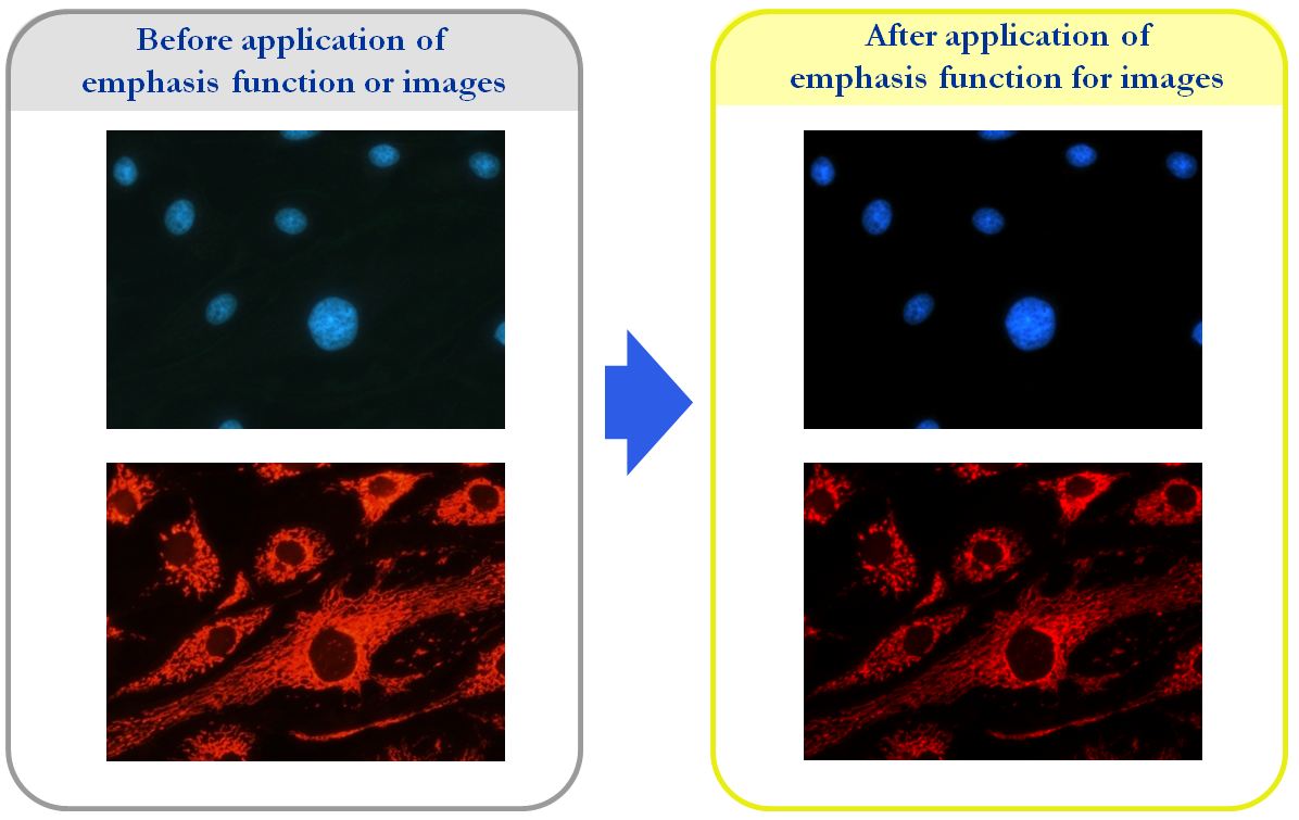
  • Channel Management – Save unique characteristics per channel

Views & Overlays

  • Well Plate Viewer
  • Color-Coded Well Plate Heat Maps

Search & Help

  • Universal Search across Recently Used items & Help topics
  • Dockable & Dynamic Help panel for easy access to Documentation

Licensing

  • Flexible License Linking/Unlinking to Computer or License Server in addition to USB dongles
  • Online License “Bundle” management – Control module configuration per base product

Data Collection & Display

  • Display Data using Heat Maps
  • Display annotated images in Data Table along with statistical data

Classification

  • Adjust measurement overlay appearances by class

Protocol-based Segmentation

  • Run on single file multi-positional (including multi-well) datasets or collections of individual images
  • Bi-direction feedback between data tables & annotated images & charts
  • Library of Protocol reports for both large datasets & single images
  • Analysis Protocols – Essentials Collection (Objects within Objects, Object Count, % Area)
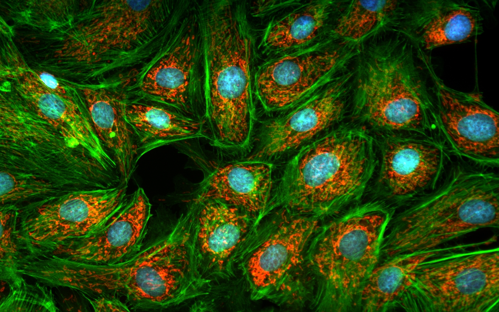
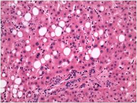
  • Analysis Protocols – Cell Biology Collection (Apoptosis, Autophagy, Cell Count, Cell Morphology & more)
  • Analysis Protocols – Cell Biology Advanced Collection (Angiogenesis, Colocalization, Neurite Outgrowth, & more)
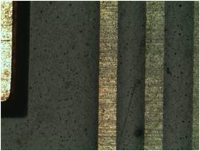
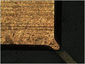
  • Analysis Protocols – Materials Collection (Particles, Pores, Particle Phase, Fiber Thickness, & more)

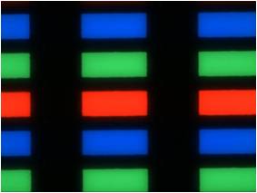
Multi-Channel Capture

  • LightSource Device Control
  • Acquisition of mixed multi-channel & transmitted light datasets
  • Automated capture of multichannel image-set (w/ a supported multi-channel light source)
  • Guided acquisition of multi-channel image-sets from a manual microscope

Real-Time Deconvolution

  • 2D Real-Time Deconvolution integrated with Capture
  • Wiener Filter-based algorithm
  • 2D Iterative-based algorithm

3D Visualization

  • 3D viewer with advanced control & volume rendering (Min & Max Projections)
  • Clipping plane viewing
  • Slicer viewing

3D Visualization of Big Data Sets

  • Convert Large Image Sets Into Pyramidal Tiff for Big Data Visualization
  • 2D Fixed & Adaptive Deconvolution Algorithms
  • Support of both Theoretical & Measured PSFs
  • GPU Acceleration
  • PSF generation for WF, CF, 2 Photon, Spinning Disk, & STED modalities
  • Spherical Abberation Correction
  • Batch Deconvolution
  • Bundles can be left as floating licenses or can be fixed to a single workstation
  • Bundle assignment to License Server can be adjusted anytime as needed
  • User time out returns license back to License Server