Product Introduction
XCAM1080PHX series cameras are designed by ToupTek as cameras with multiple output methods (HDMI+Wi-Fi+SD card), where the X in XCAM stands for multi-interface). They adopt ultra-high performance CMOS sensors. The cameras can be directly connected to HDMI displays, connected to computers and other smartphones or tablets via Wi-Fi, and can also save images and videos to SD cards for on-site analysis and subsequent research.
The greatest feature of XCAM1080PHX series cameras is that the camera itself is equipped with an embedded ARM core, which can display various camera control functions directly on HDMI displays in the form of control panels. Furthermore, users can directly control various camera parameters using a USB mouse interface mouse. Additionally, the HDMI display interface has various toolbar buttons at the bottom for user operation.
When the mouse is unplugged and a USB Wi-Fi module is inserted, and the computer is connected to that Wi-Fi, the XCAM1080PHX series camera becomes a Wi-Fi interface camera, and users can directly control the camera hardware using the included software ToupView/ToupLite. XCAM1080PHX series cameras can be used for on-site tool inspection, microscope observation, etc.


Product Features
- C-mount camera with Sony high sensitivity CMOS sensor
- HDMI / Wi-Fi can output simultaneously
- HDMI output can be achieved through built-in USB interface mouse software XCamView
- Wi-Fi output can be achieved by inserting USB interface Wi-Fi module, its control can be achieved by ToupView/ToupLite software provided with the camera
- Can achieve 5.04M pixel (2592×1944) image capture and preview (XCAM1080PHB) or 2.0M pixel (1920×1080) (XCAM1080PHD/PHE); can achieve 1080P video stream (asf format) SD card storage
- Ultra-Fine color engine ensures accurate color reproduction (when camera is connected to computer via Wi-Fi)
- Provides multi-platform standard SDK for Windows/Linux/OSX
- CNC housing one-piece design and processing
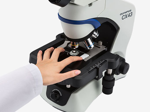
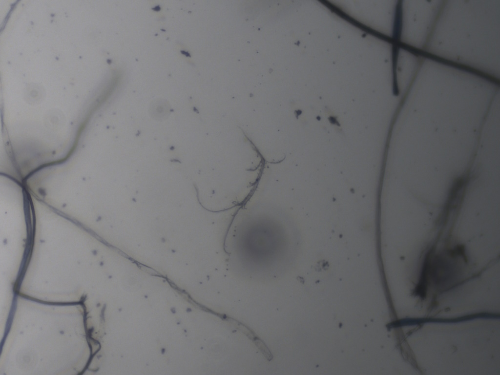
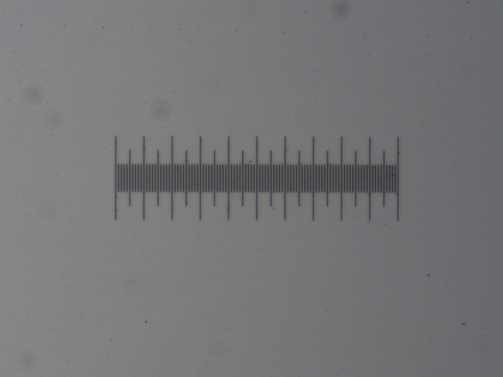
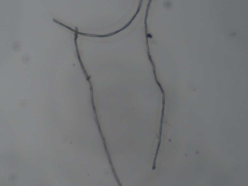
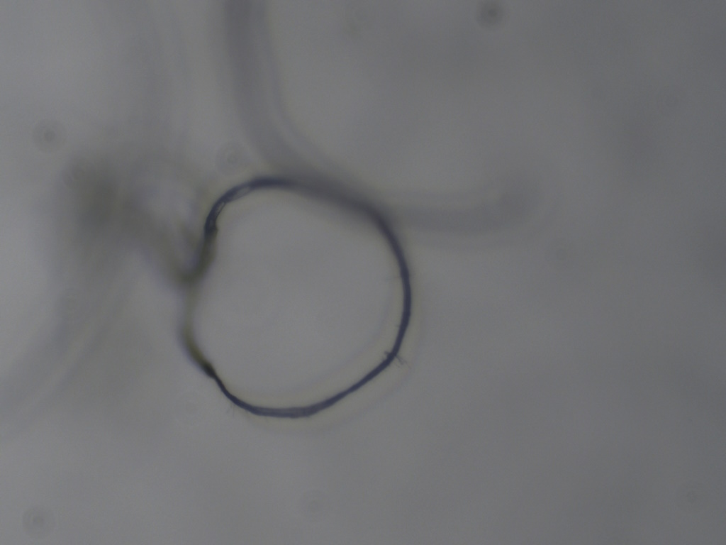
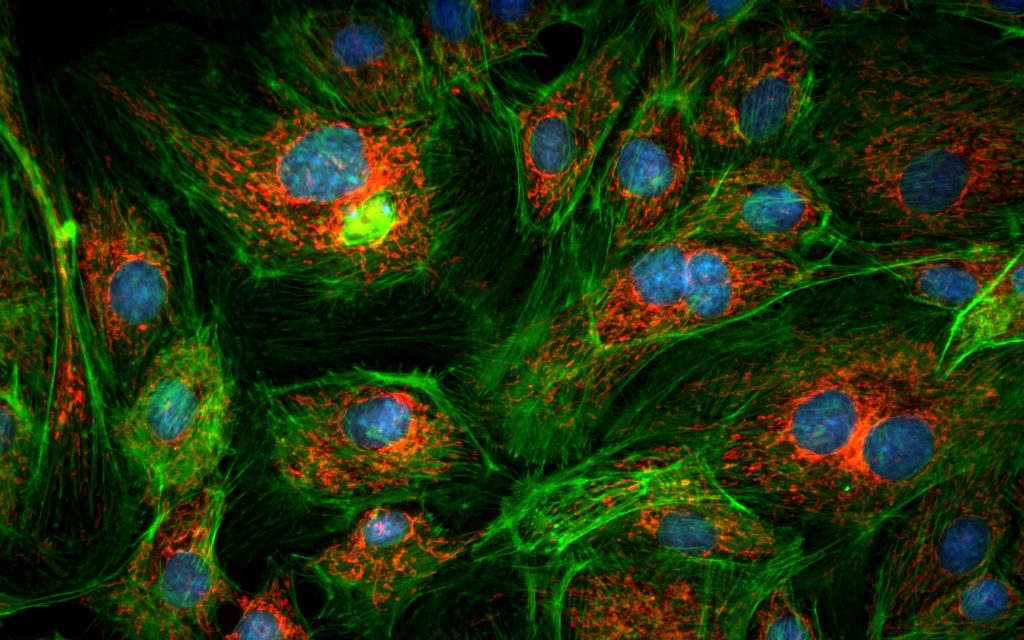
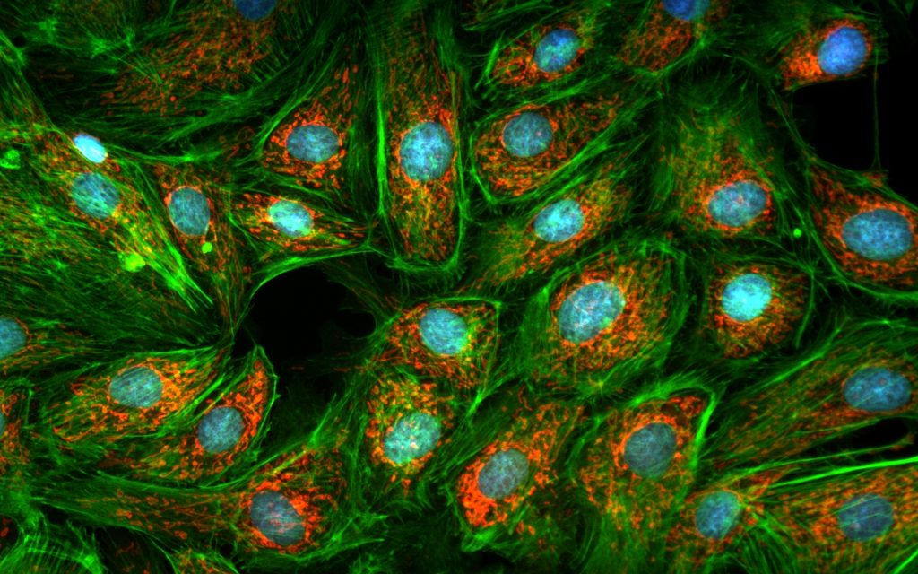
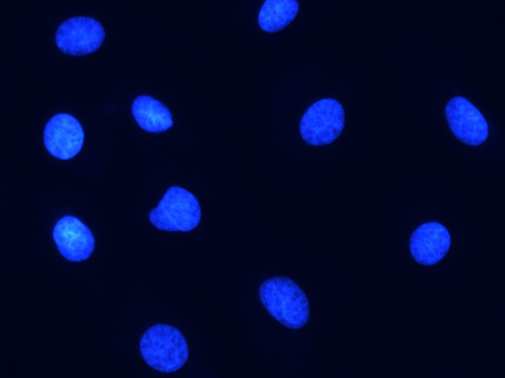
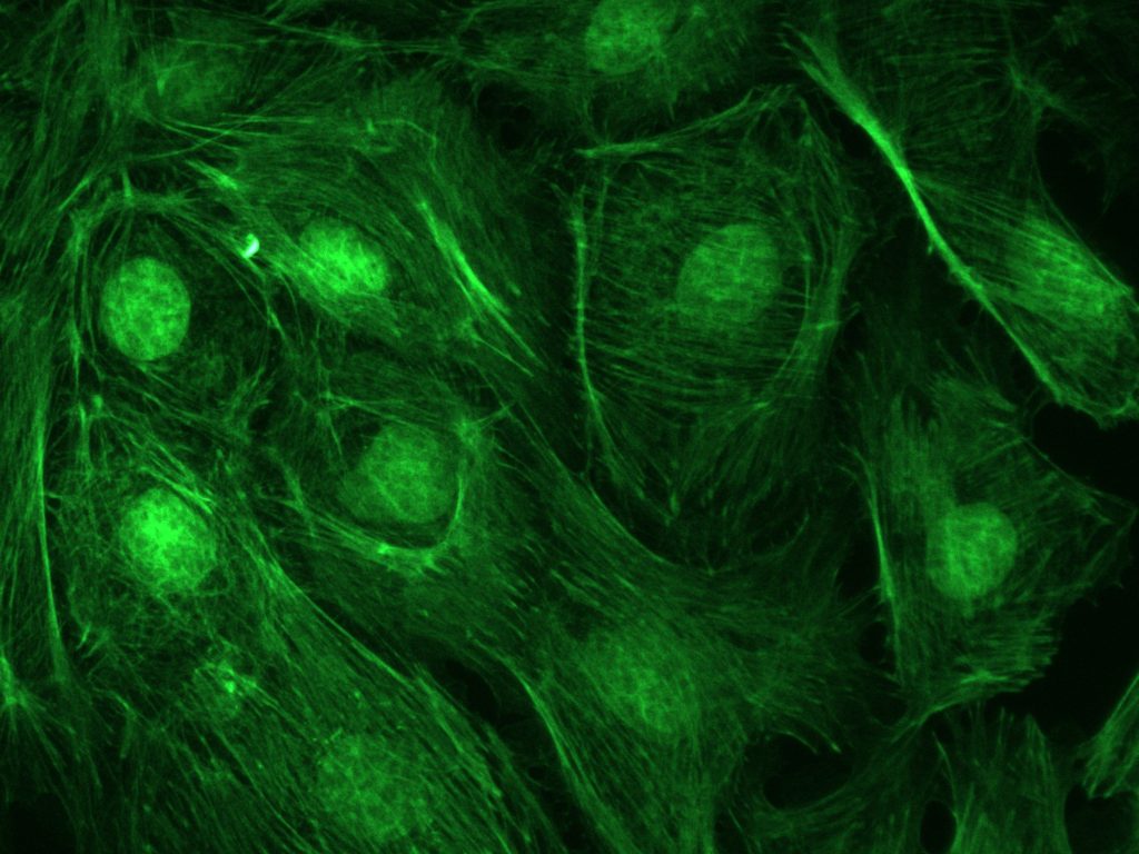
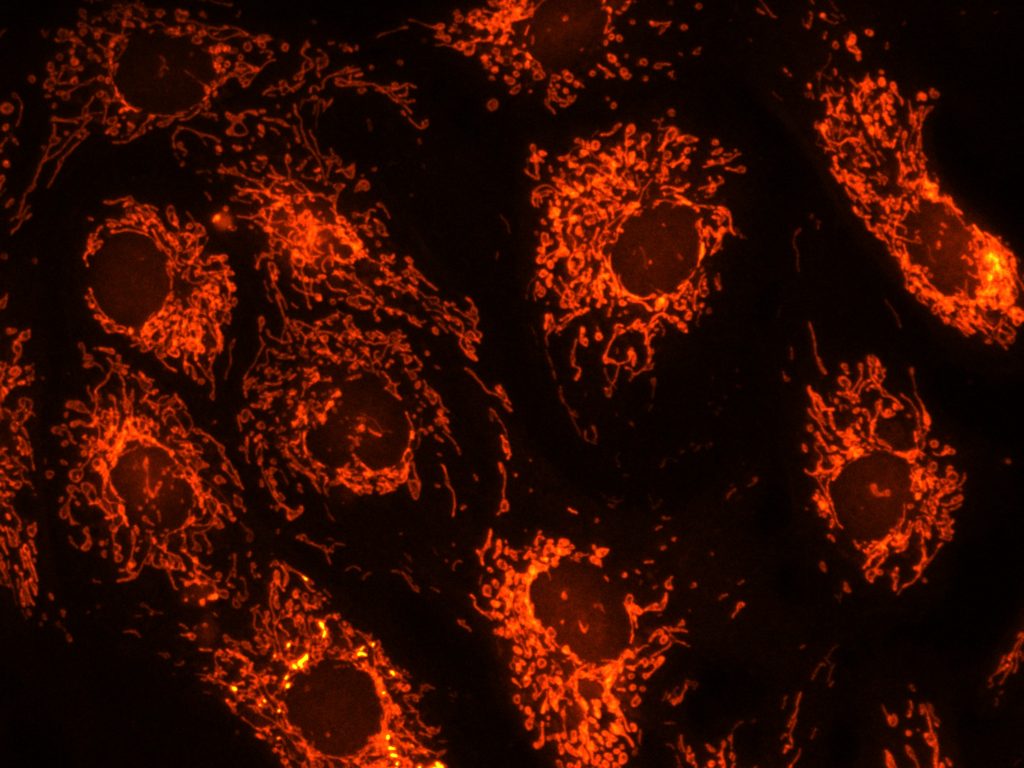
- Can be used for stereo microscope or biological microscope image acquisition, and can also be networked into interactive teaching systems
XCAM1080PHB Specifications
| Key Parameters | |
| Product Model | XCAM1080PHB |
| Product Series | C-mount HDMI+Wi-Fi CMOS camera |
| Sensor Parameters | |
| Sensor Model | Sony IMX178(C) |
| Sensor Size | 1/1.8″ (6.22×4.67 mm) |
| Diagonal | 7.8 mm |
| Pixel Size | 2.4 μm × 2.4 μm |
| Resolution | 5MP (2592×1944) |
| Shutter Type | Rolling shutter |
| Color Type | Color |
| Performance Parameters | |
| Frame Rate | HDMI: 30 fps@1920×1080; Wi-Fi: 25 fps@1920×1080 |
| Output Interfaces | HDMI, Wi-Fi |
| Lens Mount | C-mount |
| Storage Support | SD card |
| Software Support | XCamView, ToupView, ToupLite |
| Physical Parameters | |
| Power Supply | DC 12 V/1A |
| Dimensions | 78 × 70 × 92 mm |
| Weight | 0.47 kg |
| Environmental Parameters | |
| Operating Temperature | -10 °C ~ +50 °C |
| Other Information | |
| OS Support | Windows, Linux, macOS, Android, iOS |
| Certification | CE, FCC, RoHS |
Packing List

- A Camera box: 25.5 cm × 17.0 cm × 9.0 cm (1 pcs, 1.43 kg per box)
- B XCAM1080PHX series camera ×1
- C Power adapter: Input AC 100~240 V 50/60 Hz, output DC 12 V 1 A; US GS12U12-P1I (UL/CUL/BSMI/CB/FCC), EU GS12E12-P1I (TUV(GS)/CB/CE/ROHS); EMI: EN55022, EN61204-3, EN61000-3-2/-3, FCC Part 15B, BSMI CNS14338; EMS: EN61000-4-2/3/4/5/6/8/11, EN61204-3 Class A
- D HDMI cable
- E USB mouse
- F USB wireless network adapter
- G CD (drivers and application, Ø12 cm)
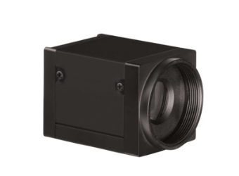
XCAM1080PHX Multi-Interface Camera
Body dimensions and interface layout

Package: 25.5 cm × 17.0 cm × 9.0 cm
- Shipping weight: approx. 1.57 kg (with packaging)
- Power: DC 12 V / 1 A (GS12 series adapter)
- Interfaces: USB 3.0 / HDMI 2.0 / LAN / WiFi (AP/STA)